. 2012 May 18;287(21):17343-17352.
doi: 10.1074/jbc.M111.314898. Epub 2012 Feb 21.

p14(ARF)-induced apoptosis in p53 protein-deficient cells is mediated by BH3-only protein-independent derepression of Bak protein through down-regulation of Mcl-1 and Bcl-xL proteins

Affiliations

p14(ARF)-induced apoptosis in p53 protein-deficient cells is mediated by BH3-only protein-independent derepression of Bak protein through down-regulation of Mcl-1 and Bcl-xL proteins

Annika Müer et al. J Biol Chem. .

Abstract

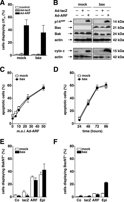
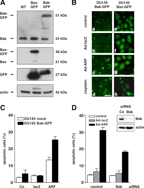
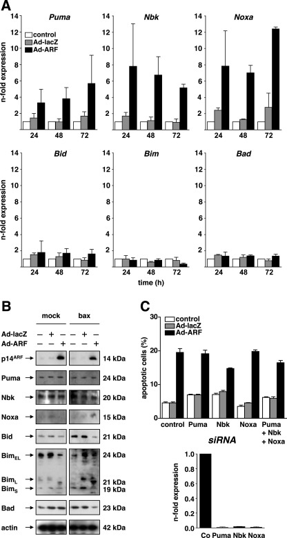
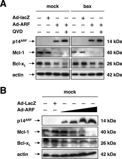
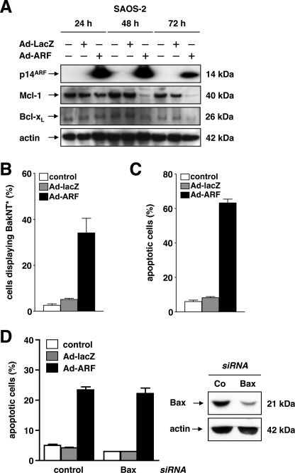
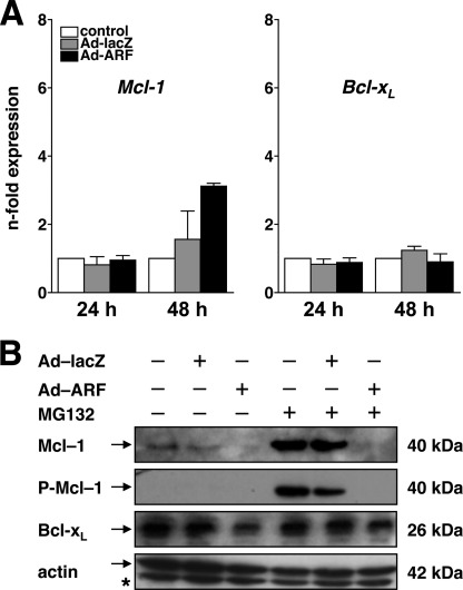
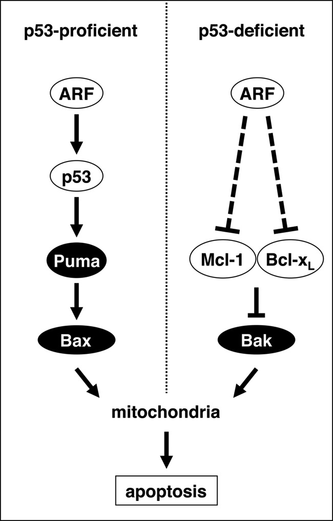
The p14(ARF) tumor suppressor plays a central role in regulating cell cycle arrest and apoptosis. We reported previously that p14(ARF) is capable of triggering apoptosis in a p53-independent manner. However, the mechanism remained unclear. Here we demonstrate that the p53-independent activation of the mitochondrial apoptosis pathway by p14(ARF) is primarily mediated by the pro-apoptotic Bax-homolog Bak. Expression of p14(ARF) exclusively triggers a N-terminal conformational switch of Bak, but not Bax, which allows for mitochondrial permeability shift, release of cytochrome c, activation of caspases, and subsequent fragmentation of genomic DNA. Although forced expression of Bak markedly sensitizes toward p14(ARF)-induced apoptosis, re-expression of Bax has no effect. Vice versa, knockdown of Bak by RNA interference attenuates p14(ARF)-induced apoptosis, whereas down-regulation of Bax has no effect. Bak activation coincides with a prominent, caspase-independent deprivation of the endogenous Bak inhibitors Mcl-1 and Bcl-x(L). In turn, mitochondrial apoptosis is fully blocked by overexpression of either Mcl-1 or Bcl-x(L). Taken together, these data indicate that in the absence of functional p53 and Bax, p14(ARF) triggers mitochondrial apoptosis signaling by activating Bak, which is facilitated by down-regulating anti-apoptotic Mcl-1 and Bcl-x(L). Moreover, our data suggest that the simultaneous inhibition of two central endogenous Bak inhibitors, i.e. Mcl-1 and Bcl-x(L), may be sufficient to activate mitochondrial apoptosis in the absence of BH3-only protein regulation.


Figures

FIGURE 1.
FIGURE 1.
P14ARF-induced cell death is Bax-independent and coincides with activation of Bak. A, p53-mutated, Bax-deficient (mock) or Bax-reconstituted (bax) DU145 cells were infected with the indicated adenoviral vectors at an m.o.i. of 50 or mock-treated for 48 h. Loss of the mitochondrial membrane potential (ΔΨm) was determined by flow-cytometry using the cationic dye 5,5′,6,6′-tetrachloro-1,1′,3,3′-tetraethyl-benzimidazolylcarbocyanin iodide. The relative number of cells displaying ΔΨm is given. B, DU145 cells were infected with the indicated adenoviral vectors (50 m.o.i.) or mock-treated. Total cellular proteins (25 μg/lane) were separated by SDS-PAGE and subjected to Western blot analysis using the indicated antibodies. C and D, cells were infected with Ad-p14ARF or mock-treated for 72 h. Apoptosis was determined by flow cytometric detection of fragmented nuclear DNA. Cells displaying a sub-G1 DNA content were less than 10% upon infection with an Ad-lacZ control adenovirus (50 m.o.i., data not shown). E and F, DU145 cells were infected with Ad-ARF or Ad-lacZ (50 m.o.i.) or mock-treated (Co) for 48 h. In parallel, cells were exposed to epirubicine (Epi, 1 μg/μl) as a positive control. An N-terminal conformational change of Bax (BaxNT*) (E) or Bak (BakNT*) (F) was studied by flow cytometry using conformation-specific antibodies. Error bars represent mean ± S.D. from triplicates.
FIGURE 2.
FIGURE 2.
P14ARF-induced mitochondrial apoptosis is mediated by Bak but not Bax. A, DU145 cells stably expressing GFP-tagged Bax or GFP-tagged Bak were generated as described (30). Total cellular proteins (25 μg/lane) were separated by SDS-PAGE and subjected to Western blot analysis using the indicated antibodies. B, DU145 Bax-GFP (left column, a–d) or Bak-GFP (right column, e–h) were either mock-treated (control) or infected with Ad-ARF or Ad-lacZ (both 50 m.o.i.). Treatment with cisplatin (10 μg/ml) was used as a positive control. The distribution of GFP-Bak and GFP-Bax was determined by fluorescence microscopy. In contrast to Bax, Bak shows a more reticular localization because of its association with mitochondria. In apoptotic cells, Bak (c and d) or Bax (h) coalesce into large clusters. Representative high-power fields at a ×400 magnification are shown. Scale bar = 50 μm). C, control vector (mock) or Bak overexpressing DU145 cells were infected with Ad-p14ARF or Ad-lacZ (both 50 m.o.i.) or mock-treated (Co), and apoptotic DNA-fragmentation was assessed by flow cytometry at 72 h after infection. D, expression of Bak was down-regulated by RNA interference (insert, confirmation by Western blot analysis). 24 h later, cells were infected with the indicated adenoviral vectors (50 m.o.i.) or treated mock for 48 h. Apoptotic DNA fragmentation was determined by flow cytometry. Error bars represent mean ± S.D. from triplicates.
FIGURE 3.
FIGURE 3.
Expression of p14ARF induces Noxa, Puma, and Nbk mRNA expression. A, DU145 cells were infected with Ad-p14ARF at an m.o.i. of 50. In parallel, cells were mock-treated (control) or infected with a control vector (Ad-lacZ, 50 m.o.i.). Cells were harvested at 24, 48, or 72 h after infection, and mRNA levels of the indicated BH3-only proteins were analyzed by quantitative PCR. B, DU145 prostate carcinoma cells were infected with the indicated adenoviral vectors (50 m.o.i.) or mock-treated for 48 h. Total cellular proteins (25 μg/lane) were separated by SDS-PAGE and subjected to Western blot analysis using the indicated antibodies. C, upper panel, DU145 cells were transfected with the indicated siRNA and cultured for 24 h. Thereafter, cells were infected with either Ad-ARF or Ad-lacZ (both 50 m.o.i.) or mock-treated (control) for 48 h. Apoptosis was assessed by flow cytometric detection of fragmented nuclear DNA. Lower panel, cells treated as described above and mRNA levels of the indicated BH3-only proteins were analyzed by quantitative PCR using cells transfected with control siRNA as reference. Error bars represent mean ± S.D. from triplicates.
FIGURE 4.
FIGURE 4.
Expression of p14ARF triggers down-regulation of anti-apoptotic Mcl-1 and Bcl-xL. A, DU145 cells were either mock-treated or infected with the indicated adenoviral vectors (50 m.o.i.) in the presence or absence of the broad-spectrum caspase inhibitor Q-VD-OPh (final concentration 10 μm) for 48 h. Total cellular proteins (25 μg/lane) were separated by SDS-PAGE and subjected to Western blot analysis using the indicated antibodies. B, dose response. Cells were infected with Ad-ARF (10, 25, 50, 100 m.o.i.). In parallel, cells were either mock-treated or infected with an Ad-lacZ (100 m.o.i.). Total cellular proteins (25 μg/lane) were subjected to SDS-PAGE and Western blot analysis with the indicated antibodies.
FIGURE 5.
FIGURE 5.
Expression of p14ARF in p53-deficient SAOS-2 cells triggers Mcl-1 and Bcl-xL deprivation and apoptosis. A, p53-deficient SAOS-2 osteosarcoma cells were infected with indicated adenoviral vectors at 50 m.o.i. or mock-treated (control). Total cellular proteins were separated by SDS-PAGE and subjected to Western blot analysis after 24, 48, and 72 h. B, SAOS-2 cells were treated as described in A. An N-terminal conformational change of Bak (BakNT*) was detected by flow cytometry using a conformation-specific antibody at 48 h after infection. C, cells were treated as described in A. Apoptotic cells were analyzed by flow cytometry to detect fragmented nuclear DNA 72 h after infection. D, expression of Bax was down-regulated by RNA interference (right panel, confirmation by Western blot analysis). 24 h later, cells were infected with the indicated adenoviral vectors (50 m.o.i.) or mock-treated for 48 h. Apoptotic DNA fragmentation was determined by flow cytometry. Error bars represent the means ± S.D. from triplicates.
FIGURE 6.
FIGURE 6.
Overexpression of Mcl-1 or Bcl-xL attenuates p14ARF-induced apoptosis. A, an pRTS1-Mcl-1 vector was stably introduced into DU145 cells to allow for the conditional expression of Mcl-1 in the presence of tetracycline (doxycycline, Dox). Cells were infected with indicated adenoviral vectors at 50 m.o.i. or mock-treated (Co). B, DU145 cells stably overexpressing Bcl-xL were infected with Ad-ARF or Ad-lacZ (both 50 m.o.i.) or mock-treated in parallel with empty vector-transduced controls (mock). Apoptosis was measured by flow cytometric detection of fragmented nuclear DNA at 72 h. The relative number of apoptotic cells is given. Error bars represent mean ± S.D. from triplicates.
FIGURE 7.
FIGURE 7.
Regulation of Mcl-1 and Bcl-xL. A, DU145 cells were infected with Ad-p14ARF or Ad-lacZ (both 50 m.o.i.) or mock-treated (control). Cells were harvested at 24 h or 48 h after infection, and mRNA expression of Mcl-1 (left panel) or Bcl-xL (right panel) was analyzed by quantitative real-time PCR. B, DU145 cells were infected with the indicated adenoviral vectors (50 m.o.i.) or mock-treated for 24 h. Thereafter, cells were cultured in the presence or absence of the proteasome inhibitor MG132 (final concentration 10 μm). Total cellular proteins were separated by SDS-PAGE and subjected to Western blot analysis using the indicated antibodies. Error bars represent the mean ± S.D. from triplicates.
FIGURE 8.
FIGURE 8.
Schematic model. Expression of the p14ARF tumor suppressor triggers both p53-dependent (left panel) and p53-independent (right panel) apoptosis signaling cascades. In p53-proficient cells, p14ARF induces the physical stabilization of p53 and the consecutive up-regulation of the BH3-only protein Puma (22). Bax and Bak then trigger the permeabilization of the outer mitochondrial membrane, the release of cytochrome c, and the activation of caspases such as caspase-9 and caspase-3/7. In p53-deficient cells, p14ARF triggers mitochondrial apoptosis via down-regulation of the anti-apoptotic Bcl-2 homologs Mcl-1 and Bcl-xL. This attenuates their inhibitory effect on pro-apoptotic Bak, which then undergoes an N-terminal conformational change to allow for the activation of mitochondria in the absence of BH3-only protein induction.

References

    1. Mao L., Merlo A., Bedi G., Shapiro G. I., Edwards C. D., Rollins B. J., Sidransky D. (1995) A novel p16INK4A transcript. Cancer Res. 55, 2995–2997 - PubMed
    1. Duro D., Bernard O., Della Valle V., Berger R., Larsen C. J. (1995) A new type of p16INK4/MTS1 gene transcript expressed in B-cell malignancies. Oncogene 11, 21–29 - PubMed
    1. Quelle D. E., Zindy F., Ashmun R. A., Sherr C. J. (1995) Alternative reading frames of the INK4a tumor suppressor gene encode two unrelated proteins capable of inducing cell cycle arrest. Cell 83, 993–1000 - PubMed
    1. Stone S., Jiang P., Dayananth P., Tavtigian S. V., Katcher H., Parry D., Peters G., Kamb A. (1995) Complex structure and regulation of the P16 (MTS1) locus. Cancer Res. 55, 2988–2994 - PubMed
    1. Zindy F., Eischen C. M., Randle D. H., Kamijo T., Cleveland J. L., Sherr C. J., Roussel M. F. (1998) Myc signaling via the ARF tumor suppressor regulates p53-dependent apoptosis and immortalization. Genes Dev. 12, 2424–2433 - PMC - PubMed

MeSH terms

Substances