. 2012 Apr;32(8):1337-53.
doi: 10.1128/MCB.06525-11. Epub 2012 Feb 21.

Determinants for activation of the atypical AGC kinase Greatwall during M phase entry

Affiliations

Determinants for activation of the atypical AGC kinase Greatwall during M phase entry

Kristina A Blake-Hodek et al. Mol Cell Biol. 2012 Apr.

Abstract

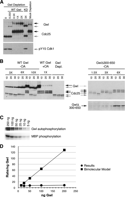
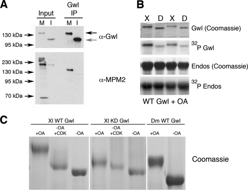
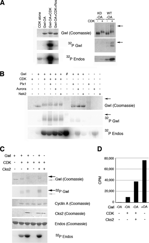
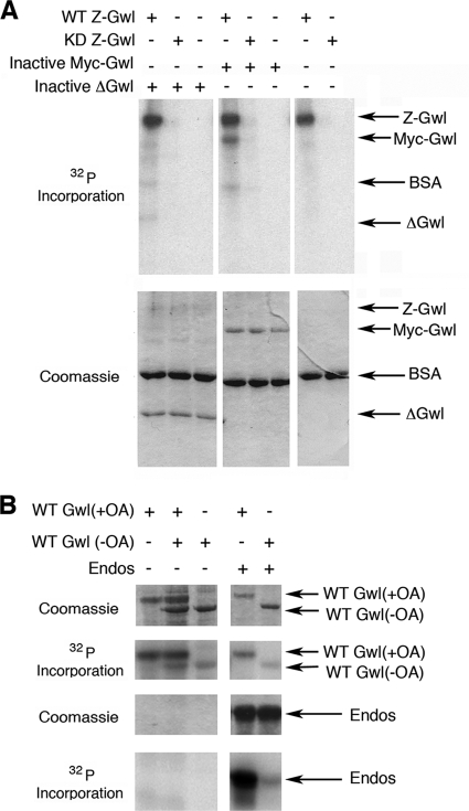
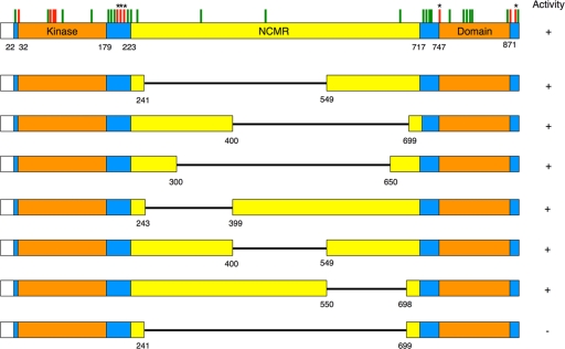
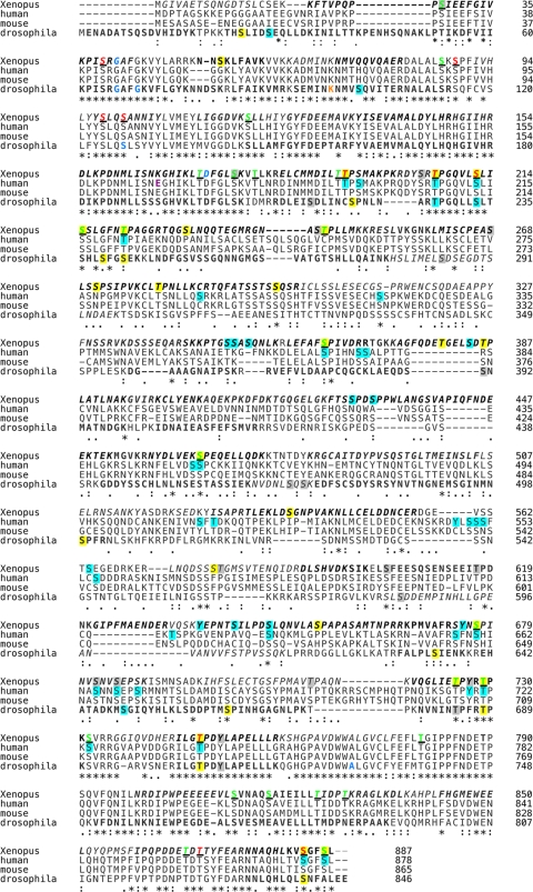
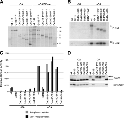
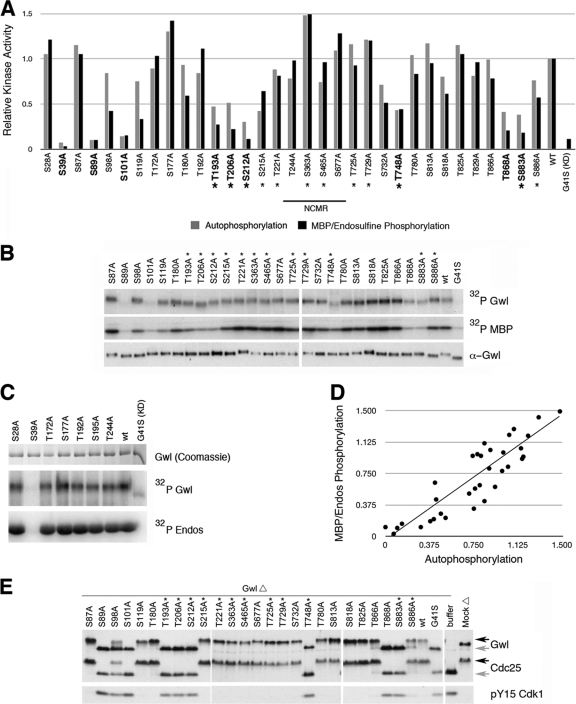
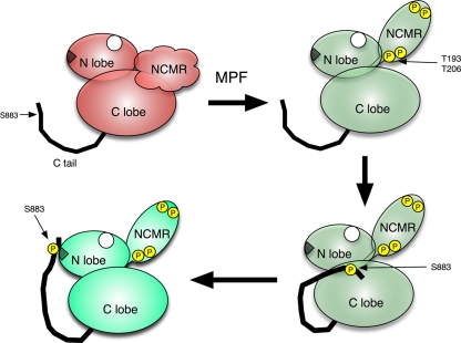
The atypical AGC kinase Greatwall (Gwl) mediates a pathway that prevents the precocious removal of phosphorylations added to target proteins by M phase-promoting factor (MPF); Gwl is thus essential for M phase entry and maintenance. Gwl itself is activated by M phase-specific phosphorylations that are investigated here. Many phosphorylations are nonessential, being located within a long nonconserved region, any part of which can be deleted without effect. Using mass spectrometry and mutagenesis, we have identified 3 phosphorylation sites (phosphosites) critical to Gwl activation (pT193, pT206, and pS883 in Xenopus laevis) located in evolutionarily conserved domains that differentiate Gwl from related kinases. We propose a model in which the initiating event for Gwl activation is phosphorylation by MPF of the proline-directed sites T193 and T206 in the presumptive activation loop. After this priming step, Gwl can intramolecularly phosphorylate its C-terminal tail at pS883; this site probably plays a role similar to that of the tail/Z motif of other AGC kinases. These events largely (but not completely) explain the full activation of Gwl at M phase.


Figures

Fig 1
Fig 1
Assays for Gwl kinase activity and biological function. (A and B) Extract assays. CSF extracts were Gwl or mock depleted and then supplemented with WT or KD Gwl (from OA-treated Sf9 cells) or with buffer. 0.25X etc. indicate the concentrations of exogenous Gwl relative to normal endogenous levels. After 30 min at 25°C, the extracts were analyzed for Gwl, Cdc25C, and Tyr15 (pY15) on Cdk1. M phase Gwl and Cdc25 (black arrows) migrate more slowly than their interphase counterparts (gray arrows). (A) WT Gwl at a 0.5× concentration maintains depleted extracts in M phase, but KD Gwl cannot prevent M phase exit even at a 3× concentration. (B) Left, Gwl-depleted CSF extracts were supplemented with Gwl proteins or buffer alone (Gwl Depl). After 10, 20, and 30 min of incubation, the extracts were analyzed as described for panel A; here, Cdc25 is a doublet. WT Gwl made in the absence of OA restores M phase only at a 6× concentration and only after 30 min (presumably after cyclin accumulation). Right, Gwl-depleted extracts were supplemented with Gwl with a deletion of residues 300 to 650 (see Fig. 4) made in the absence of OA. The Gwl(Δ300-650) enzyme is more easily activated than the WT by the MPF activity that briefly remains immediately after depletion (45). (C) Standard kinase assays. Both autophosphorylation and phosphorylation of the model substrate MBP are linear from 5 ng to 250 ng of WT Gwl made in the presence of OA. (D) Gwl autophosphorylation is a unimolecular reaction. The initial rate of autophosphorylation (from the experiment whose results are shown in panel C; circles) is independent of Gwl concentration. Squares show expectations if the reaction was instead bimolecular (between two Gwl molecules or Gwl and a contaminating kinase).
Fig 2
Fig 2
Gwl phosphorylation. (A) M phase but not interphase Gwl carries an MPM-2 epitope. CSF extracts were directly analyzed (M) or treated with calcium (interphase extracts; I). Input lanes contain 10% of the extract subjected to Gwl immunoprecipitation (IP). (B) Kinase activities of WT Xenopus (X) and Drosophila (D) Gwl (made in OA-treated Sf9 cells). Kinase assays contained 10 ng (left lanes) or 5 ng of Gwl (right lanes). The autophosphorylation activity of the fly enzyme is 30 to 40% of that of frog Gwl, but both phosphorylate endosulfine (Endos) to similar extents. (C) Coomassie blue staining of samples analyzed by MS in the experiment whose results are reported in Table 1. KD Gwl in OA-treated Sf9 cells is less phosphorylated than WT Gwl because the KD enzyme does not autophosphorylate. In vitro activation of WT interphase Gwl (made in the absence of OA) by Cdk2/cyclin A produces a smear of hyperphosphorylated Gwl. Fly Gwl (Dm) is 41 amino acids shorter than frog Gwl (Xl).
Fig 3
Fig 3
Activation of Gwl by CDKs. (A) Left, 250 ng of WT Gwl made in Sf9 cells without OA was treated with 50 ng of Cdk2/cyclin A as indicated. In the far-right lane, the CDK inhibitor roscovitine (Ros) was added at 0.25 mM. One-tenth of each reaction mixture was added to a standard kinase assay to monitor Gwl autophosphorylation or phosphorylation of endosulfine. Right, the results of a similar experiment using 50 ng of MPF (Cdk1/cyclin B). Roscovitine was added to each standard kinase assay to inactivate the MPF. To resolve phosphorylated Gwl, the gel in the right panel was run longer than that at the left; arrows indicate the position of active Gwl (made in the presence of OA) in a separate lane. (B) Possible roles of other M phase-specific kinases in activating Gwl were tested. In the tabulation at the top, plus signs (+) indicate 250 ng of WT inactive Gwl (made in the absence of OA) and/or 50 ng of the other indicated kinases, and the pound sign indicates 50 ng of active WT Gwl made in the presence of OA (arrows). (C and D) The Suc1 paralog Cks2 enhances CDK-driven activation of Gwl. Cks2 was added to reaction mixtures containing Cdk2/cyclin A and Gwl made in the absence of OA. In the results shown in panel D (measured on phosphocellulose filters), Cks2 enhanced CDK's activation of Gwl from 12.5% to 49% of the specific activity of Gwl made in the presence of OA. Endos, endosulfine.
Fig 4
Fig 4
Gwl autophosphorylation is primarily intramolecular. (A) Amounts of 25 ng of WT or KD Gwl (both containing a Z tag and made in OA-treated Sf9 cells) were incubated in kinase assays with 250 ng of either heat-inactivated Gwl(Δ300-650) or heat-inactivated Myc-tagged WT Gwl (the Myc tag is smaller than the Z tag) as indicated. Despite the 10× molar excess of the heat-inactivated substrates, most 32P incorporation was into the active WT Z-tagged enzyme. (B) WT Gwl made in the presence or absence of OA was quantified by Coomassie blue staining (top). Bottom, 100 ng of active Gwl and/or 250 ng of inactive Gwl was added as indicated to standard kinase reactions that contained endosulfine (Endos) or no substrate. The active enzyme [Gwl(+OA)] did not phosphorylate the inactive one [Gwl(−OA)].
Fig 5
Fig 5
Schematic view of Gwl structure and mutant analysis. In WT Xenopus Gwl, the two halves of the conserved kinase domain are orange, the NCMR is yellow, and blue shows regions that distinguish Gwl from other AGC kinases. In the top row, red tick marks indicate S/T-to-A mutants with lower function than the WT, while green tick marks indicate mutants with near-normal activity. Asterisks (*) mark sites essential for Gwl function whose M phase phosphorylation has been MS verified. In subsequent rows, black lines indicate deleted parts of the NCMR. Except for Δ241-699, all these deletions exhibit near-wild-type activities in kinase and CSF extract rescue assays (see Fig. 7; also data not shown).
Fig 6
Fig 6
Gwl phosphorylation sites and mutagenesis. Xenopus Gwl aligned with human, mouse, and Drosophila counterparts. In vivo mitotic Xenopus Gwl was purified by immunoprecipitation from CSF extracts or by expression in OA-treated Sf9 insect cells with nearly identical results; in vitro Gwl was made in Sf9 cells in the absence of OA and then treated in vitro with CDKs. Amino acids covered by MS analysis with both in vivo and in vitro Gwl are in boldface italics; those found only with in vivo enzymes are in boldface; those found only in the in vitro Gwl are in italics; and regions of Gwl not detected in any MS studies are in normal font. Yellow (in vivo and in vitro), aqua (in vivo only), or gray (in vitro only) shading indicates phosphorylated S, T, or Y residues identified here or in published studies of human Gwl (7, 8, 28). Underlined letters represent S/T-to-A mutations made in frog Gwl. Mutations that disrupt Gwl function in extracts are shown in red; mutations without effect are in green. Blue letters indicate sites of known loss-of-function mutations in fly (G66S, G69E, S127L, and A728V) or frog (G41S and D173A) Gwl (44, 45).
Fig 7
Fig 7
NCMR deletions. (A) WT, KD, and deletion mutant Gwl were purified from Sf9 cells in the presence or absence of OA. Proteins were stained with Coomassie blue; those purified from OA-treated cells were treated with λ phosphate (PPase) before electrophoresis because hyperphosphorylated Gwl runs as a smear. Numbers in parentheses indicate concentrations relative to that of the WT. (B) Kinase activities of Gwl deletion mutants. Amounts of 50 ng of the indicated Gwl proteins were added to standard kinase assays to monitor both autophosphorylation and the labeling of myelin basic protein (MBP). (C) Graphic representation of the experimental results shown in panel B. The y axis depicts relative specific kinase activities (that is, kinase activity per nanogram of Gwl) normalized with respect to the specific activity of WT Gwl made in the presence of OA. (D) Function of Gwl deletions in egg extracts. CSF extracts were immunodepleted for Gwl and then supplemented with recombinant Gwl at the original endogenous levels. Extracts were incubated at 25°C for 30 min before immunoblotting. Cdc25C becomes hyperphosphorylated in M phase (black arrow; the gray arrow indicates the interphase form), while Y15 of Cdk1 is phosphorylated during interphase but not M phase.
Fig 8
Fig 8
Analysis of S/T-to-A mutations. (A) Summary showing specific kinase activities of S/T-to-A mutants expressed in OA-treated cells. Sites marked with asterisks were observed by MS to be phosphorylated in M phase. Sites in boldface are required for Gwl function in extracts (see panel E). Normalization to the specific activity of WT Gwl was as described for Fig. 7C. (B and C) Kinase assays. Amounts of 50 ng of the indicated proteins were assayed in vitro for autophosphorylation and for kinase activity against MBP (B) or endosulfine (Endos) as described for Fig. 7B. Gwl was quantified by Western blot assay (B) or by Coomassie blue staining after λ protein phosphatase treatment (C). (D) Relationship between autophosphorylation and phosphorylation of exogenous substrates, based on the results shown in panels B and C. Numbers on each axis represent the relative specific activities of mutant proteins (with OA) relative to that of WT Gwl (with OA); each point represents the results for one mutant protein. (E) Functional analysis of S/T-to-A mutant proteins in CSF extracts. Physiological activities of the mutant proteins were determined as described for Fig. 7D; concentrations of exogenous recombinant Gwl proteins were equivalent to that of endogenous Gwl.
Fig 9
Fig 9
Phosphomimetic mutations. (A to C) S/T residues of importance were mutated to D/E, and the proteins expressed in OA-treated Sf9 cells. Mutants were assayed for kinase activity (A and B) and function in CSF extracts (C) as described for Fig. 7. (D to F) Phosphomimetic Gwl mutants with limited constitutive activity, made in Sf9 cells without OA. (D and E) Standard kinase assays with 5 to 20 ng of mutant Gwl. In the experiment whose results are shown in panel E, 2 ng of active WT Gwl (+OA) was the control. S101D and S101D T193E mutants made in the absence of OA displayed 3% and 15%, respectively, of the control's specific activity, even though the mutants remain hypophosphorylated (in panel E, the arrow indicates active M phase Gwl). (F) Biological assays of S101D T193E double phosphomimetic mutant Gwl made in the absence of OA. This protein rescues M phase in Gwl-depleted extracts, but only at 4× excess.
Fig 10
Fig 10
Kinases targeting key phosphosites in Gwl. (A) The indicated Gwl fragments were expressed as glutathione S-transferase fusions in Escherichia coli and then treated with MPF or Plx1 in the presence of 32P ATP in standard kinase assays. The fragment containing residues 185 to 213 contains two proline-directed phosphosites (T193 and T206). The results of T-to-A mutations show that both sites are targeted by MPF, although T193 is the more efficient substrate. MPF does not target the S883-containing fragment. Plx1 does not phosphorylate any of these Gwl fragments, though it is active against Pin1, a previously demonstrated substrate (9). (B) Gwl but not Plx1 or CDK can phosphorylate a peptide containing S883. The C-terminal peptide RRNNAQHLKVSGFSL (CT) was incubated with WT or KD Gwl (made in the presence of OA) or with Plx1 (PL) or Cdk1/cyclin A (CD) in standard kinase assays. Incorporation of 32P into the peptide (in counts per minute) was measured (see Materials and Methods). The results of two independent experiments are shown; black and gray bars show the results of independent trials of each experiment. Activity of the Plx1 preparation is demonstrated by phosphorylation of a PLKtide peptide substrate (PT).
Fig 11
Fig 11
Gwl is an atypical AGC kinase. Alignment contains primary sequences of Xenopus Gwl and human PKCθ, PKBα, and PKA. Aqua highlighting shows residues in Gwl that are conserved in other enzymes. Gray highlighting shows residues shared by two or more AGC kinases but not Gwl. Yellow highlighting shows residues found in Gwl and other AGC kinases not depicted (PKBβ, PKCα, PKCβ, PKCι, PKCζ, or PDK1). The red-shaded site is the site phosphorylated by PDK1 in most AGC activation domains; to aid orientation, this single site is shown on both sides of the NCMR. Red dots indicate phosphorylation sites in the C-terminal tails of AGC kinases that commonly participate in activation. Underlined amino acids in Gwl were mutated; underlined red letters show S/T-to-A changes that significantly reduce Gwl activity, while underlined green amino acids are nonessential. Structural annotations are from references (brown) and (purple and orange).
Fig 12
Fig 12
Homology structural modeling. (A) Xenopus Gwl (red) modeled relative to human PKA (blue). The Gwl structure lacks the first 25 N-terminal amino acids and the NCMR, which have no equivalents in PKA. Gwl's C-terminal tail (“C” in red) is shorter than PKA's (“C” in blue) and thus cannot extend as far. The arrow points to PKA's turn motif. Alpha helices B and C (α-Β and α-C) demarcate two sides of the pocket in the N-terminal lobe (boxed region) that normally binds the hydrophobic domain (HF) of AGC kinases. (B) Close-up of the boxed region in panel A, showing the binding of PKA's C-terminal HF (green) to its corresponding pocket, which stabilizes the active configuration of α-C. Residues within the pocket important for HF interactions are labeled. (C) Close-up of the region of Gwl in the box in panel A. Gwl's N-terminal lobe has residues likely to form an HF binding pocket, but the C-terminal tail is too short to contact this pocket intramolecularly.
Fig 13
Fig 13
Gwl proteins with phosphomimetic mutations of S883 do not exhibit constitutive activity in vivo. mRNAs for FLAG-tagged S883E or S883D Gwl were injected into immature G2 Xenopus oocytes that were treated (or not) with progesterone (43). (A) Untreated injected oocytes remained immature (IM), with Gwl in its interphase form; expression of S883E or S883D did not induce germinal vesicle breakdown (data not shown). Germinal vesicle breakdown was induced in ∼100% of oocytes treated with progesterone (GV); Gwl acquired M phase phosphorylations. (B and C) Gwl was immunoprecipitated from injected oocytes and subjected to standard kinase assays with (B and C) or without (C) MBP substrate. S883D and S883E Gwl proteins made in immature interphase oocytes have no intrinsic constitutive activity, but they are strongly activated during progesterone-induced maturation.
Fig 14
Fig 14
A model for Gwl activation. Gwl is depicted with four domains: the N-terminal lobe (N lobe), the C-terminal lobe (C lobe), the nonconserved middle region (NCMR), and the C-terminal tail (thick black line). During M phase entry, MPF phosphorylates Gwl at the presumptive activation loop sites T193 and T206. MPF, and perhaps other kinases, may also target several phosphosites within the NCMR that could have redundant functions. NCMR phosphorylations could conceivably change its conformation to allow substrates access to Gwl's active site between the N- and C-terminal lobes. After Gwl is primed by MPF, S883 in the C-terminal tail can be autophosphorylated at the active site. It is likely that pS883 can subsequently interact with a patch of basic residues in the N lobe (dark triangle) to help stabilize active Gwl (40). The round hole in the N lobe depicts the conserved HF binding pocket (Fig. 12), whose role is not yet clear.

Comment in

Similar articles

Cited by

References

    1. Adams JA. 2003. Activation loop phosphorylation and catalysis in protein kinases: is there functional evidence for the autoinhibitor model? Biochemistry 42: 601–607 - PubMed
    1. Archambault V, Zhao X, White-Cooper H, Carpenter AT, Glover DM. 2007. Mutations in Drosophila Greatwall/Scant reveal its roles in mitosis and meiosis and interdependence with Polo kinase. PLoS Genet. 3: e200. - PMC - PubMed
    1. Behn-Krappa A, Newton AC. 1999. The hydrophobic phosphorylation motif of conventional protein kinase C is regulated by autophosphorylation. Curr. Biol. 9: 728–737 - PubMed
    1. Burgess A, et al. 2010. Loss of human Greatwall results in G2 arrest and multiple mitotic defects due to deregulation of the cyclin B-Cdc2/PP2A balance. Proc. Natl. Acad. Sci. U. S. A. 107: 12564–12569 - PMC - PubMed
    1. Castilho PV, Williams BC, Mochida S, Zhao Y, Goldberg ML. 2009. The M phase kinase Greatwall (Gwl) promotes inactivation of PP2A/B55delta, a phosphatase directed against CDK phosphosites. Mol. Biol. Cell 20: 4777–4789 - PMC - PubMed

Publication types

LinkOut - more resources