. 2012 May;32(9):1604-17.
doi: 10.1128/MCB.06547-11. Epub 2012 Feb 21.

A balance between Tel1 and Rif2 activities regulates nucleolytic processing and elongation at telomeres

Affiliations

A balance between Tel1 and Rif2 activities regulates nucleolytic processing and elongation at telomeres

Marina Martina et al. Mol Cell Biol. 2012 May.

Abstract

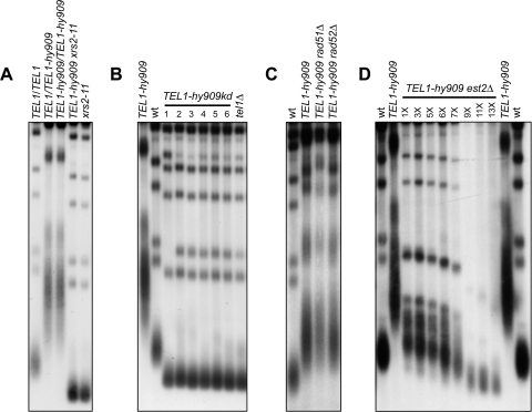
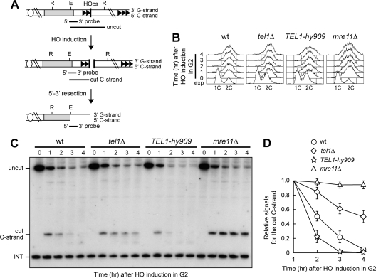
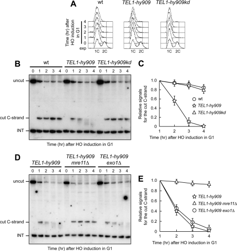
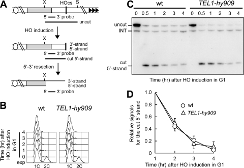
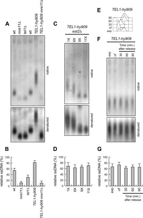
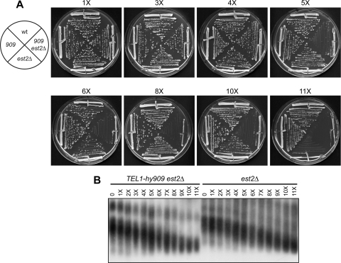
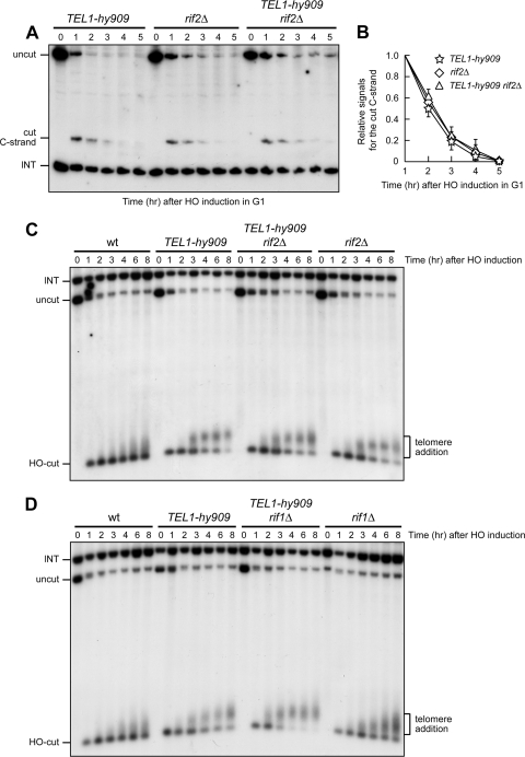
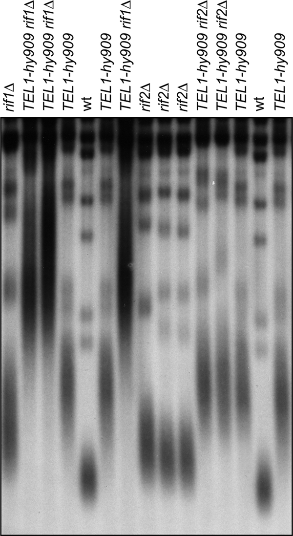
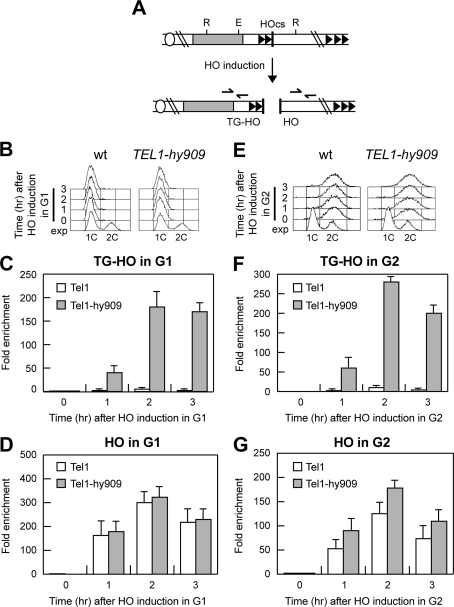
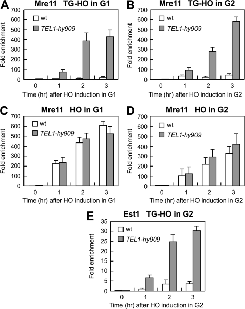
Generation of G-strand overhangs at Saccharomyces cerevisiae yeast telomeres depends primarily on the MRX (Mre11-Rad50-Xrs2) complex, which is also necessary to maintain telomere length by recruiting the Tel1 kinase. MRX physically interacts with Rif2, which inhibits both resection and elongation of telomeres. We provide evidence that regulation of telomere processing and elongation relies on a balance between Tel1 and Rif2 activities. Tel1 regulates telomere nucleolytic processing by promoting MRX activity. In fact, the lack of Tel1 impairs MRX-dependent telomere resection, which is instead enhanced by the Tel1-hy909 mutant variant, which causes telomerase-dependent telomere overelongation. The Tel1-hy909 variant is more robustly associated than wild-type Tel1 to double-strand-break (DSB) ends carrying telomeric repeat sequences. Furthermore, it increases the persistence at a DSB adjacent to telomeric repeats of both MRX and Est1, which in turn likely account for the increased telomere resection and elongation in TEL1-hy909 cells. Strikingly, Rif2 is unable to negatively regulate processing and lengthening at TEL1-hy909 telomeres, indicating that the Tel1-hy909 variant overcomes the inhibitory activity exerted by Rif2 on MRX. Altogether, these findings highlight a primary role of Tel1 in overcoming Rif2-dependent negative regulation of MRX activity in telomere resection and elongation.


Figures

Fig 1
Fig 1
Telomere overelongation in TEL1-hy909 cells. (A to C) XhoI-cut genomic DNA from exponentially growing cells (YEPD at 25°C) was subjected to Southern blot analysis using a radiolabeled poly(GT) telomere-specific probe. For panel B, 6 independent TEL1-hy909kd transformant clones were analyzed (lanes 1 to 6). (D) Meiotic tetrads from EST2/est2Δ TEL1/TEL1-hy909 diploid cells were dissected on YEPD plates. After ∼25 generations, TEL1-hy909 est2Δ spore clones were streaked for successive times (1 to 13 times) and aliquots of cells from the indicated streaks were propagated in YEPD liquid medium for 5 h to prepare genomic DNA for telomere length detection, as in panels A to C. Each subsequent streak represents ∼25 generations of growth. wt, wild type.
Fig 2
Fig 2
Effects of the tel1Δ and TEL1-hy909 mutations on ssDNA generation at a DSB end carrying telomeric repeats. (A) Schematic representation of the HO cleavage site (HOcs) with the TG repeat sequences (81 bp; two arrowheads) that were placed centromere proximal to the HO site at the ADH4 locus on chromosome VII-L. The centromere is shown as a circle on the left. The probe used to monitor nucleolytic degradation of the 5′ C strand is also indicated. R, RsaI; E, EcoRV. (B to D) HO expression was induced at time zero by galactose addition to nocodazole-arrested cell cultures that were kept arrested in G2. (B) Fluorescence-activated cell sorter analysis of DNA content. exp, exponentially growing cells. (C) RsaI- and EcoRV-digested genomic DNA was hybridized with a single-stranded riboprobe that anneals to the 5′ C strand to a site located 212 bp from the HO cutting site. The probe reveals an uncut 390-nt DNA fragment (uncut), which is converted by HO cleavage into a 166-nt fragment (cut C strand). Degradation of the 5′ C strand leads to disappearance of the probe signal as resection proceeds beyond the hybridization region. The probe also detects a 138-nt fragment from the ade2-101 locus on chromosome XV (INT), which serves as internal loading control. (D) Densitometric analysis. Plotted values are means ± SDs from three independent experiments, as in panel C.
Fig 3
Fig 3
Tel1-hy909 allows resection in G1 of a DSB adjacent to telomeric repeats. (A to C) HO expression was induced at time zero by galactose addition to α-factor-arrested cells, all carrying the system described in Fig. 2A. Cells were kept arrested in G1. (A) Fluorescence-activated cell sorter analysis of DNA content. (B) RsaI- and EcoRV-digested genomic DNA was analyzed as described in the legend to Fig. 2C. (C) Densitometric analysis. Plotted values are means ± SDs from three independent experiments, as in panel B. (D and E) HO expression was induced at time zero by galactose addition to α-factor-arrested cells that were kept arrested in G1. Cell cycle arrest was verified by fluorescence-activated cell sorter analysis (data not shown). (D) RsaI- and EcoRV-digested genomic DNA was analyzed as described in the legend to Fig. 2C. (E) Densitometric analysis. Plotted values are means ± SDs from three independent experiments, as in panel D.
Fig 4
Fig 4
Tel1-hy909 does not enhance nucleolytic processing in G1 of a DSB with no telomeric repeats. (A) Schematic representation of the system used to generate the HO-induced DSB on chromosome VII. SspI- and XbaI-digested genomic DNA is hybridized with the indicated single-stranded probe that anneals to the 5′ strand to a site located 248 nt from the HO cutting site, revealing an uncut 897-nt DNA fragment (uncut), which is converted by HO cleavage into a 286-nt fragment (cut 5′ strand). Loss of the 5′ strand beyond the hybridization region of the probe leads to disappearance of the signal generated by the probe. S, SspI; X, XbaI. (B to D) HO expression was induced at time zero by galactose addition to α-factor-arrested wild-type and TEL1-hy909 cells, all carrying the system depicted in panel A. Cells were kept arrested in G1. (B) Fluorescence-activated cell sorter analysis of DNA content. (C) SspI- and XbaI-digested genomic DNA was hybridized with the probe described in panel A. (D) Densitometric analysis. Plotted values are means ± SDs from four independent experiments, as in panel C.
Fig 5
Fig 5
Effects of tel1Δ and TEL1-hy909 alleles on ssDNA formation at native telomeres. (A) Genomic DNA prepared from exponentially growing cells was digested with XhoI, and single-stranded G tails were visualized by in-gel hybridization (native) using an end-labeled C-rich oligonucleotide as a probe. The gel was then denatured and hybridized again with the same probe for loading control (denatured). (B) The amount of native TG-ssDNA, as in panel A, was normalized to the total amount of TG sequences detected in each denatured sample. (C and D) Meiotic tetrads from EST2/est2Δ TEL1/tel1-hy909 diploid cells were dissected on YEPD plates. (C) After ∼25 generations, TEL1-hy909 est2Δ spore clones were streaked for successive times (1 to 11 times), and aliquots of cells from the indicated streaks were propagated in YEPD liquid medium for 5 h to prepare genomic DNA for telomeric ssDNA detection, as in panel A. (D) The amount of native TG-ssDNA, as in panel C, was normalized to the total amount of TG sequences detected in each denatured sample. (E to G) Exponentially growing (exp) TEL1-hy909 cells were arrested in G1 with α-factor (αf) and released into the cell cycle. (E) Fluorescence-activated cell sorter analysis of DNA content. (F) Genomic DNA prepared at the indicated time points after release from the α-factor block was analyzed for telomeric ssDNA detection, as in panel A. (G) The amount of native TG-ssDNA, as in panel F, was normalized to the total amount of TG sequences detected in each denatured sample. Plotted values in panels B, D, and G are means ± SDs from three independent experiments.
Fig 6
Fig 6
The TEL1-hy909 mutation accelerates the onset of senescence in est2Δ cells. Meiotic tetrads from an EST2/est2Δ TEL1/tel1-hy909 diploid strain were dissected on YEPD plates. (A) After ∼25 generations, spore clones from 15 tetrads were streaked for successive times (1 to 11 times). (B) An aliquot of cells from the indicated streaks was propagated in YEPD liquid medium for 5 h to prepare genomic DNA for telomere length determination by Southern blot analysis. All tetratype tetrads behaved as the one shown in panel A.
Fig 7
Fig 7
Processing and elongation of a DSB end carrying TG tracts in TEL1-hy909 cells are not affected by RIF2 deletion. (A and B) HO expression was induced at time zero by galactose addition to α-factor-arrested cells, all carrying the system described in Fig. 2A. Cells were kept arrested in G1, and cell cycle arrest was verified by fluorescence-activated cell sorter analysis (data not shown). (A) RsaI- and EcoRV-digested genomic DNA was analyzed as described in the legend to Fig. 2C. (B) Densitometric analysis. Plotted values are means ± SDs from three independent experiments, as in panel A. (C and D) HO expression was induced at time zero by galactose addition to exponentially growing cells. AvaI- and NdeI-digested genomic DNA was subjected to Southern blot analysis using a TRP1 probe, which reveals the ∼800-bp AvaI-HO fragment exposing the TG repeats, whose length progressively increases as new telomere repeats are added. A bracket points out new telomere repeats added to the exposed TG-HO sequence.
Fig 8
Fig 8
Different effects of RIF1 and RIF2 deletion on the length of native TEL1-hy909 telomeres. XhoI-cut genomic DNA from exponentially growing cell cultures with the indicated genotypes was analyzed as described in the legend to Fig. 1.
Fig 9
Fig 9
Association of Tel1-HA and Tel1-hy909-HA to DNA ends. (A) The system described in Fig. 2A was used to generate TG-HO and HO DNA ends, and the primers used to detect protein association centromere proximal (TG-HO) or centromere distal (HO) to the HO-induced DSB are represented by arrows in the bottom part of the panel. (B to G) HO expression was induced at time zero by galactose addition to G1-arrested (B to D) or G2-arrested (E to G) TEL1-HA (wild-type) and TEL1-hy909-HA (TEL1-hy909) cells, which were kept arrested in G1 or G2 by α-factor and nocodazole, respectively. (B and E) Fluorescence-activated cell sorter analysis of DNA content. (C to G) Chromatin samples taken at the indicated times after HO induction were immunoprecipitated with anti-HA antibody. (C and F) Coimmunoprecipitated DNA was analyzed by qPCR using primer pairs located 640 bp centromere proximal to the HO cleavage site (HOcs; TG-HO) and at the nontelomeric ARO1 fragment of chromosome IV (CON). (D and G) Coimmunoprecipitated DNA was analyzed by qPCR using primer pairs located 550 bp centromere distal to the HO cleavage site (HOcs; HO) and at the nontelomeric ARO1 fragment of chromosome IV (CON). In all graphs, data are expressed as relative fold enrichment of the TG-HO or HO signal over the CON signal after normalization to input signals for each primer set. The data presented are means ± SDs from three different experiments.
Fig 10
Fig 10
Mre11 and Est1 association to DNA ends in wild-type and TEL1-hy909 cells. G1- or G2-arrested wild-type and TEL1-hy909 cells carrying the TG-HO system described in Fig. 2A and expressing fully functional MYC-tagged Mre11 (A to D) or MYC-tagged Est1 (E) were treated as described in the legend to Fig. 9, and cell cycle arrest was verified by fluorescence-activated cell sorter analysis (data not shown). Chromatin samples taken at the indicated times after HO induction were immunoprecipitated with anti-MYC antibody. (A, B, and E) Coimmunoprecipitated DNA was analyzed by qPCR with the primer pairs used for Fig. 9C and F. (C and D) Coimmunoprecipitated DNA was analyzed by qPCR with the primer pairs used for Fig. 9D and G. In all graphs, data are expressed as relative fold enrichment of the TG-HO or HO signal over the CON signal after normalization to input signals for each primer set. The data presented are means ± SDs from three different experiments.

References

    1. Abdallah P, et al. 2009. A two-step model for senescence triggered by a single critically short telomere. Nat. Cell Biol. 11:988–993 - PMC - PubMed
    1. Arnerić M, Lingner J. 2007. Tel1 kinase and subtelomere-bound Tbf1 mediate preferential elongation of short telomeres by telomerase in yeast. EMBO Rep. 8:1080–1085 - PMC - PubMed
    1. Baldo V, Testoni V, Lucchini G, Longhese MP. 2008. Dominant TEL1-hy mutations compensate for Mec1 lack of functions in the DNA damage response. Mol. Cell. Biol. 28:358–375 - PMC - PubMed
    1. Bianchi A, Shore D. 2007. Increased association of telomerase with short telomeres in yeast. Genes Dev. 21:1726–1730 - PMC - PubMed
    1. Bianchi A, Negrini S, Shore D. 2004. Delivery of yeast telomerase to a DNA break depends on the recruitment functions of Cdc13 and Est1. Mol. Cell 16:139–146 - PubMed

Publication types

MeSH terms

Substances