. 2012 May;32(9):1733-44.
doi: 10.1128/MCB.06717-11. Epub 2012 Feb 21.

Rab GTPase-activating proteins in autophagy: regulation of endocytic and autophagy pathways by direct binding to human ATG8 modifiers

Affiliations

Rab GTPase-activating proteins in autophagy: regulation of endocytic and autophagy pathways by direct binding to human ATG8 modifiers

Doris Popovic et al. Mol Cell Biol. 2012 May.

Abstract

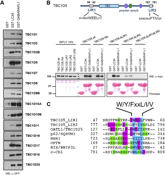
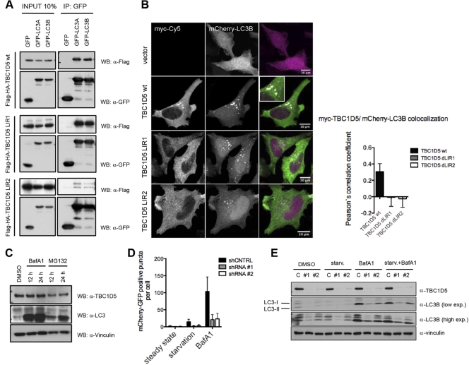
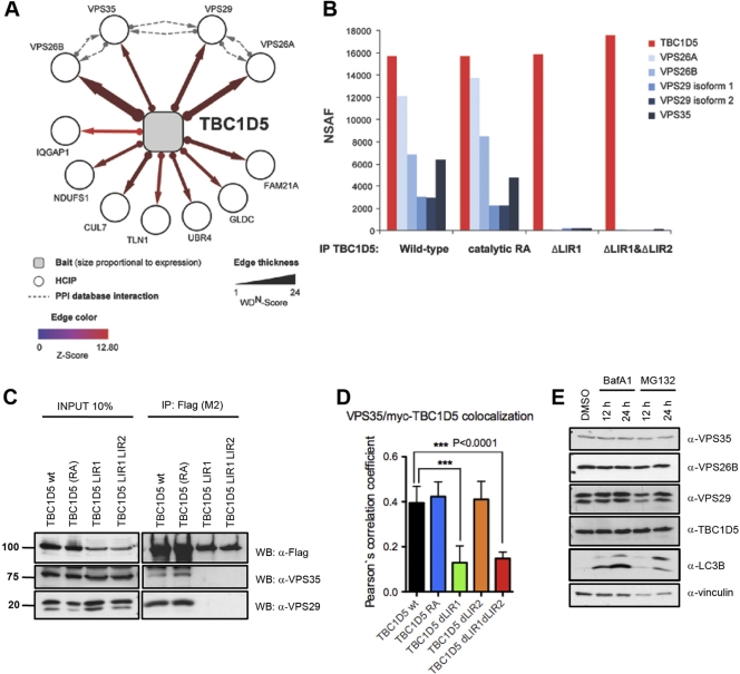
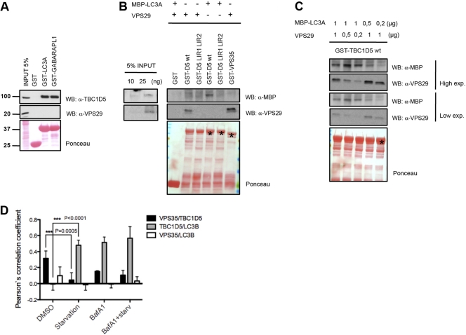
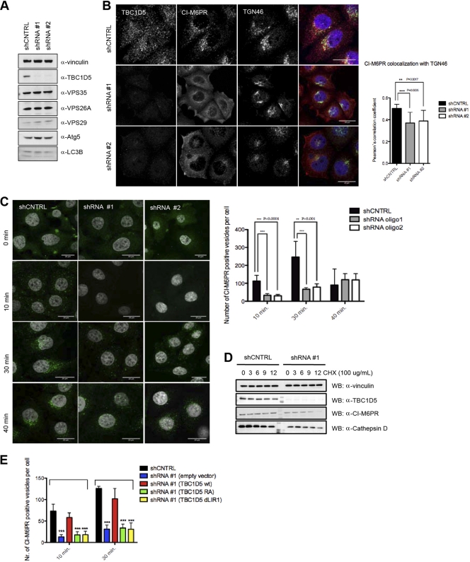
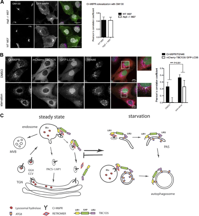
Autophagy is an evolutionarily conserved degradation pathway characterized by dynamic rearrangement of membranes that sequester cytoplasm, protein aggregates, organelles, and pathogens for delivery to the vacuole and lysosome, respectively. The ability of autophagosomal membranes to act selectively toward specific cargo is dependent on the small ubiquitin-like modifier ATG8/LC3 and the LC3-interacting region (LIR) present in autophagy receptors. Here, we describe a comprehensive protein-protein interaction analysis of TBC (Tre2, Bub2, and Cdc16) domain-containing Rab GTPase-activating proteins (GAPs) as potential autophagy adaptors. We identified 14 TBC domain-containing Rab GAPs that bind directly to ATG8 modifiers and that colocalize with LC3-positive autophagy membranes in cells. Intriguingly, one of our screening hits, TBC1D5, contains two LIR motifs. The N-terminal LIR was critical for interaction with the retromer complex and transport of cargo. Direct binding of the retromer component VPS29 to TBC1D5 could be titrated out by LC3, indicating a molecular switch between endosomes and autophagy. Moreover, TBC1D5 could bridge the endosome and autophagosome via its C-terminal LIR motif. During starvation-induced autophagy, TBC1D5 was relocalized from endosomal localization to the LC3-positive autophagosomes. We propose that LC3-interacting Rab GAPs are implicated in the reprogramming of the endocytic trafficking events under starvation-induced autophagy.


Figures

Fig 1
Fig 1
TBC proteins bind human ATG8 proteins via canonical LIR. (A) ATG8-interacting TBC proteins. HEK 293T cell lysates overexpressing GFP-tagged human TBC proteins were subjected to pulldown assays using GST, GST-MAP1LC3A, and GST-GABARAPL1. Coprecipitated TBC proteins were detected with anti-GFP antibody. Inputs are shown in Fig. S1A in the supplemental material. WB, Western blot. (B) (Top) Schematic representation of TBC1D5 with Pfam domains annotated and LIR motifs identified. (Bottom) The wild type and catalytic RA and LIR mutants of TBC1D5 were transiently expressed in 293T cells and subjected to pulldown assays with GST alone or GST-ATG8 proteins. GST fusion proteins were visualized by Ponceau S staining. (C) Alignments of canonical LIR motifs from annotated ATG8-interacting partners and LIR motifs mapped in TBC1D5. Magenta, acidic residues; blue, small and hydrophobic residues; green, serine residues.
Fig 2
Fig 2
TBC1D5 is implicated in regulation of autophagy flux. (A) Coimmunoprecipitation (IP) of wild-type TBC1D5 and the ΔLIR1 and ΔLIR2 mutants with MAP1LC3A and MAP1LC3B proteins transiently overexpressed in 293T cells. (B) Immunofluorescence assay of myc-tagged TBC1D5 (wild type and ΔLIR1 and ΔLIR2 mutants) and mCherry-MAP1LC3B transiently overexpressed in HeLa cells. HeLa cells were cultured in regular medium, washed with PBS, fixed in 2% paraformaldehyde (PFA), and incubated with primary antibodies against myc tag. (C) Abundance of endogenous TBC1D5 and MAP1LC3 in HeLa cells 12 and 24 h after treatment with BafA1 (200 nM) and MG132 (10 μM), respectively. The indicated proteins were detected in whole-cell lysates by immunoblotting. DMSO, dimethyl sulfoxide. (D) shRNA control (shCNTRL) and stable knockdown cell lines expressing tandem mCherry-GFP-MAP1LC3B were subjected to starvation conditions or treated with BafA1 for 4 h. Autophagosomes were quantified in more than 100 cells in 3 independent experiments. A paired t test was used to indicate significance. The error bars represent standard deviations (SD) (GraphPad Prism). (E) shRNA control cells and two stable TBC1D5 knockdown HeLa cell lines were starved (starv.) in EBSS, treated with bafilomycin A1, or starved in the presence of BafA1 (4 h). Total cell lysates were analyzed by SDS-PAGE following immunoblotting of endogenous TBC1D5, MAP1LC3B, and vinculin (as a loading control). exp., expression.
Fig 3
Fig 3
TBC1D5 requires LIR1 to bind to the retromer. (A) Proteomic analysis of TBC1D5. The association map shows identified HCIPs. The dashed lines indicate interactions annotated in the BIOGRID, MINT, and HPRD protein interaction databases. (B) Proteomic analysis of TBC1D5. Immune complexes from cells expressing wild-type and catalytic RA and ΔLIR mutant TBC1D5 proteins were subjected to IP–LC–MS-MS. Normalized spectral abundance factors (NSAF) were calculated based on total spectral counts (41). (C) Validation of TBC1D5-associated proteins. Expression of HA-Flag-tagged wild-type TBC1D5, TBC1D5 RA, TBC1D5 ΔLIR1, or TBC1D5 ΔLIR1 ΔLIR2 was induced in stable TREx293 cells for 24 h. Cell lysates were immunoprecipitated with Flag M2-coupled resin and immunoblotted with anti-Flag and anti-Vps29 antibody, respectively. Units for numbers (left) are kDa. (D) Immunofluorescence of endogenous Vps35 and transiently expressed myc-tagged TBC1D5 (wild type [wt], TBC1D5 RA, TBC1D5 ΔLIR1, TBC1D5 ΔLIR2, and TBC1D5 ΔLIR1 ΔLIR2) in HeLa cells. Colocalization of more than 100 cells was quantified in 3 independent experiments for each TBC1D5 construct using Volocity Demo software, and a paired t test was performed using GraphPad Prism. The error bars indicate SD. (E) Abundance of endogenous TBC1D5, MAP1LC3, and retromer subunits VPS35, VPS26B, and VPS29 in HeLa cells 12 and 24 h after treatment with BafA1 (100 nM) and MG132 (10 μM).
Fig 4
Fig 4
TBC1D5 partitions between LC3 and retromer-mediated functions. (A) Lysates from nontransfected and HA-Flag-VPS35-, HA-Flag-VPS26A-, and HA-Flag-VPS26B-transfected 293T cells were incubated with GST, GST-MAP1LC3A, and GST-GABARAPL1. Copurifying proteins were detected by immunoblotting with anti-Flag, anti-VPS29, and anti-TBC1D5 antibodies. Units for numbers (left) are kDa. (B) In vitro binding assay with purified VPS29, MBP-coupled MAP1LC3A, and GST-TBC1D5 fusion protein. MAP1LC3A or VPS29 (1 μg) was used in a reaction volume of 400 μl (right). (C) In vitro competition assay. Concentrations of VPS29 and MAP1LC3A are indicated. GST-fused proteins TBC1D5, TBC1D5 ΔLIR1 ΔLIR2, and VPS35 are indicated by the asterisk on the Ponceau stain. (D) Immunofluorescence colocalization quantification of a double-stable U2OS cell line expressing mCherry-TBC1D5 and GFP-LC3B under normal conditions and starvation or bafilomycin A1 and starvation plus BafA1 treatment. Endogenous VPS35 was stained, and the colocalization of all three proteins in more than 200 cells was analyzed in Volocity Demo. The data were compared using a paired t test (GraphPad). The error bars indicate SD.
Fig 5
Fig 5
TBC1D5 regulates retrograde transport and the autophagy pathway. (A) Total cell lysates from shRNA control and TBC1D5 knockdown cells were analyzed on SDS-PAGE and blotted for TBC1D5 and retromer subunits, respectively. (B) CI-M6PR localization is changed upon TBC1D5 knockdown. shRNA control cells and TBC1D5 knockdown cells were fixed and costained with primary antibodies against CI-M6PR and TGN46. Colocalization of both was analyzed in more than 200 cells in 3 independent experiments using Volocity Demo. The error bars indicate SD. (C) TBC1D5 knockdown affects CI-M6PR internalization. An antibody-feeding assay was performed as described in Materials and Methods, and CI-M6PR-positive vesicles were quantified in more than 300 cells using an ImageJ particle analysis plug-in. (D) TBC1D5 is important for stability of CI-M6PR and cathepsin D sorting. A cycloheximide (CHX) chase was performed on shRNA control and TBC1D5 knockdown cells, using 100 μg/ml of CHX and chase for 12 h. Total cell lysates were blotted against endogenous CI-M6PR, cathepsin D, and TBC1D5. (E) Rescue experiment for CI-M6PR retrieval to the TGN. shRNA control cells were subjected to an antibody-feeding assay as described for panel C; at the 10- and 30-min time points, cells were fixed, permeabilized, and incubated with primary antibody for TGN46 and then stained with secondary antibodies for both primary antibodies, CI-M6PR and TGN46. Colocalization of the internalized fraction of CI-M6PR with TGN46 was analyzed using Volocity Demo. Knockdown cells were transiently transfected with myc-tagged shRNA-resistant clones of TBC1D5, the wild type as well as RA and ΔLIR1 mutants. An antibody-feeding assay was performed 16 h posttransfection, and cells were fixed and stained using the same procedure described above. The graph presents quantification of CI-M6PR-positive vesicles quantified for more than 200 transfected cells. An unpaired t test was performed using GraphPad Prism. ***, P < 0.0005.
Fig 6
Fig 6
Autophagy and retrograde transport are interdependent. (A) Localization of GFP-CI-M6PR and Golgi marker GM130 in WT and ATG5 knockout (KO) mouse embryonic fibroblastes (MEFs) and quantification of GFP-CI-M6PR colocalization with Golgi marker GM130 in WT and ATG5 KO MEFs. Colocalization with Golgi marker GM130 was analyzed in more than 60 transfected MEFs using Volocity Demo. The error bars indicate SD. (B) Immunofluorescence analysis of localization of CI-M6PR and TGN46 in double-stable U2OS cell lines expressing mCherry-TBC1D5 and GFP-MAP1LC3B. Cells were starved for 4 h in EBSS, fixed, and stained with corresponding endogenous antibodies. Colocalization of CI-M6PR/TGN46 and TBC1D5/MAP1LC3B was analyzed on more than 200 cells using Volocity Demo, and a paired t test was performed in GraphPad Prism. (C) Working model for TBC1D5 function in retrograde transport of CI-M6PR and autophagy. In steady state, TBC1D5 is localized on endosomes via binding to the retromer complex, regulating the dynamics of retrograde transport of cargo–CI-M6PR. Upon starvation-induced autophagy, TBC1D5 binds to autophagosomes via LIR2 and is transferred to the autophagosomal membrane via LIR1 and LIR2.

Similar articles

Cited by

References

    1. Adachi A, et al. 2010. Golgi-associated GSK3beta regulates the sorting process of post-Golgi membrane trafficking. J. Cell Sci. 123:3215–3225 - PubMed
    1. Arighi CN, Hartnell LM, Aguilar RC, Haft CR, Bonifacino JS. 2004. Role of the mammalian retromer in sorting of the cation-independent mannose 6-phosphate receptor. J. Cell Biol. 165:123–133 - PMC - PubMed
    1. Attar N, Cullen PJ. 2010. The retromer complex. Adv. Enzyme Regul. 50:216–236 - PubMed
    1. Barr F, Lambright DG. 2010. Rab GEFs and GAPs. Curr. Opin. Cell Biol. 22:461–470 - PMC - PubMed
    1. Behrends C, Sowa ME, Gygi SP, Harper JW. 2010. Network organization of the human autophagy system. Nature 466:68–76 - PMC - PubMed

Publication types

MeSH terms

Substances