. 2012 May;32(9):1618-32.
doi: 10.1128/MCB.06727-11. Epub 2012 Feb 21.

Altered replication in human cells promotes DMPK (CTG)(n) · (CAG)(n) repeat instability

Affiliations

Altered replication in human cells promotes DMPK (CTG)(n) · (CAG)(n) repeat instability

Guoqi Liu et al. Mol Cell Biol. 2012 May.

Abstract

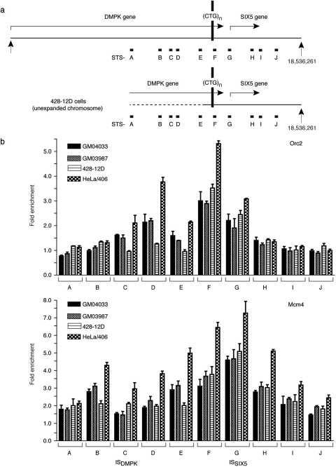
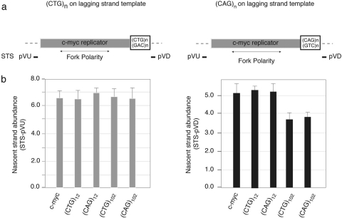
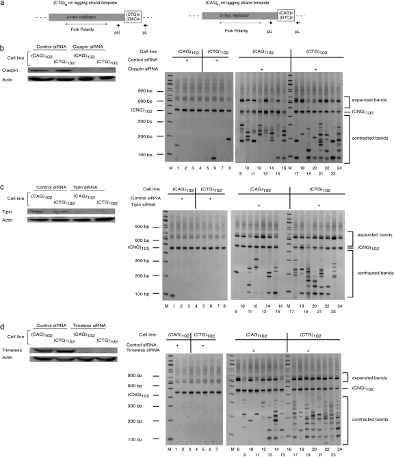
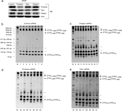
Myotonic dystrophy type 1 (DM1) is associated with expansion of (CTG)(n) · (CAG)(n) trinucleotide repeats (TNRs) in the 3' untranslated region (UTR) of the DMPK gene. Replication origins are cis-acting elements that potentiate TNR instability; therefore, we mapped replication initiation sites and prereplication complex protein binding within the ~10-kb DMPK/SIX5 locus in non-DM1 and DM1 cells. Two origins, IS(DMPK) and IS(SIX5), flanked the (CTG)(n) · (CAG)(n) TNRs in control cells and in DM1 cells. Orc2 and Mcm4 bound near each of the replication initiation sites, but a dramatic change in (CTG)(n) · (CAG)(n) replication polarity was not correlated with TNR expansion. To test whether (CTG)(n) · (CAG)(n) TNRs are cis-acting elements of instability in human cells, model cell lines were created by integration of cassettes containing the c-myc replication origin and (CTG)(n) · (CAG)(n) TNRs in HeLa cells. Replication forks were slowed by (CTG)(n) · (CAG)(n) TNRs in a length-dependent manner independent of replication polarity, implying that expanded (CTG)(n) · (CAG)(n) TNRs lead to replication stress. Consistent with this prediction, TNR instability increased in the HeLa model cells and DM1 cells upon small interfering RNA (siRNA) knockdown of the fork stabilization protein Claspin, Timeless, or Tipin. These results suggest that aberrant DNA replication and TNR instability are linked in DM1 cells.


Figures

Fig 1
Fig 1
TNR instability at the DMPK locus. (a) Structural features and map of the DMPK/SIX5 region. The bent arrows indicate the 3′ end of the DMPK gene and the transcriptional start site of the SIX5 gene (nucleotide positions are based on GenBank accession number NT_011109). The primers used for the PCR shown in panel b, PEf, PDMPK, and PSIX5, are indicated by three short horizontal arrows. DMPK, dystrophia myotonica protein kinase gene; SIX5, sine oculis homeobox homolog 5 gene; DMPK-AT, DMPK antisense transcript attenuated by CTCF binding (14). (b) Agarose gel electrophoresis of PCR products amplified by upper primer PEf or PDMPK and lower primer PSIX5 using genomic DNA from six cell types as templates. PCR bands amplified from unexpanded (CTG)n · (CAG)n (n = 8 to 27) and bands from expanded (CTG)n · (CAG)n (n = 80 to 780) are indicated by arrows. Note that the binding site for PEf has been deleted in the hemizygous cell line 428-12D (lane 12). M, molecular size markers. (c) Primer sites A, C, and E are present as single copies in 428-12D cells. Quantitative PCR was used to measure the copy number of STS-A, -C, and -E sequences in genomic DNAs. A-10, A-30, A-50, A-75, and A-100 indicate quantitation of sites ∼10, 30, 50, 75, and 100 kb upstream of STS-A in 428-12D cell genomic DNA. (d) Primer sites A, C, and E are present on the same chromosome.
Fig 2
Fig 2
Replication origin activity in the DMPK/SIX5 region in non-DM1 cells. (a) Structural features and map of the DMPK/SIX5 locus (see the legend to Fig. 1a). A to J denote the 5′ nucleotide coordinates of STSs used for real-time qPCR. (b) DNA replication initiation sites mapped by 0.5- to 2-kb nascent-DNA abundance assay at sequence-tagged sites A to J normalized against an internal-control low NDAA STS-54.8 (β-globin locus [42]). The error bars indicate SD.
Fig 3
Fig 3
Assembly of pre-RC on the DMPK/SIX5 region in non-DM1 cells. (a) Orc2 binding at the DMPK/SIX5 region. Shown is immunoprecipitation of cross-linked chromatin from asynchronous IMR90, MM, and HeLa/406 cells with antibodies against human Orc2. The immunoprecipitated DNA was used for qPCR amplification by primers at STS-A to -J. The relative enrichment (ordinate) at each STS is the copy number of the DNA precipitated with the specific antibody divided by the copy number of the DNA at the same STS precipitated by preimmune serum. (b) Binding of Mcm4 at the DMPK/SIX5 region assayed by immunoprecipitation of cross-linked chromatin, as in panel a, with antibody against human Mcm4. The immunoprecipitated DNA was used for qPCR amplified by primers at STS-A to -J, as in panel a. The error bars indicate SD.
Fig 4
Fig 4
Replication origin activity at the DMPK/SIX5 region in DM1 cell lines. DNA replication activity (nascent-strand abundance) was measured as in Fig. 2. GM04033 and GM03987 are DM1 patient fibroblasts with 1.5- to 2.0-kb TNR expansions (Fig. 1b) on one allele. 428-12D is a lymphoblastoid cell line with ∼1- to 3.5-kb TNR expansions on the expanded chromosome and a large deletion of ISDMPK upstream of the DMPK TNR on the unexpanded chromosome (Fig. 1b). HeLa cell data from Fig. 2 are included for comparison. The error bars indicate SD.
Fig 5
Fig 5
Binding of Orc2 and Mcm4 on the DMPK/SIX5 region in DM1 cells. Chromatin immunoprecipitation was performed as in Fig. 3 to assay Orc2 binding (a) and Mcm4 binding (b) in GM04033, GM03987, and 428-12D fibroblasts. HeLa cell data from Fig. 3 are included for comparison. The error bars indicate SD.
Fig 6
Fig 6
Fork stalling at an ectopic chromosomal site is TNR repeat length dependent and TNR orientation independent. (a) Diagram of HeLa/406 cell-derived isogenic cell lines created by Flp recombinase-mediated site-specific integration (56, 57). A 2.4-kb c-myc replicator (gray bar) was connected to a (CTG)n · (CAG)n (n = 12, 102 repeats) repeat tract (open bar). The positions of sequence-tagged sites STS-pVU and STS-pVD used in qPCR, in flanking vector sequences upstream and downstream of the origin, respectively, are also indicated. (b) Nascent-DNA (0.5 to 2.0 kb) abundance measured by qPCR at STS-pVU and STS-pVD in HeLa Flp recombinase-treated cell lines harboring (CTG)n · (CAG)n (n = 12, 102) sequences in either orientation relative to the c-myc replicator or the wild-type c-myc replicator (c-myc; n = 0). The error bars indicate SD.
Fig 7
Fig 7
The fork protection complex is essential for (CTG)102 · (CAG)102 stability. (a) Diagram of the ectopic c-myc–(CTG) · (CAG) integration site. pU and pL are small-pool PCR primers used to detect TNR length. (b) (Left) Immunoblot confirmation of siRNA knockdown of Claspin. Actin, loading control. (Right) spPCR of DNA from c-myc–(CAG)102 · (CTG)102 [(CAG)102] or c-myc–(CTG)102 · (CAG)102 [(CTG)102] cells treated with control siRNA or Claspin siRNA analyzed by PAGE. Similar results were obtained with other control siRNAs. M, molecular size markers. (c) (Left) Immunoblot confirmation of siRNA knockdown of Tipin. (Right) spPCR of DNA from c-myc–(CAG)102 · (CTG)102 or c-myc–(CTG)102-(CAG)102 cells treated with control siRNA or Tipin siRNA. (d) (Left) Immunoblot confirmation of siRNA knockdown of Timeless. (Right) spPCR of DNA from c-myc–(CAG)102 · (CTG)102 or c-myc–(CTG)102 · (CAG)102 cells treated with control siRNA or Timeless siRNA. Cells were treated twice, 48 h apart, with siRNA and analyzed 48 h later. The low-mobility shadow bands observed above the ∼450-bp amplification product of the (CTG)102 · (CAG)102 progenitor sequence in control cell DNA and the corresponding bands in siRNA-treated cell DNA are slipped-strand products of reannealing during the PCR (71), as demonstrated by reamplification of the purified linear ∼450-bp product band (56).
Fig 8
Fig 8
The fork protection complex is essential for DMPK (CTG)n · (CAG)n stability in DM1 cells. (a) Immunoblot (WB) confirmation of siRNA knockdown of Timeless, Tipin, or Claspin in DM1 cells (GM04033). Actin, loading control. (b, c, d, and e) spPCR of DNA from cells treated as in Fig. 7 with control siRNA (lanes 1 to 8), Claspin siRNA (lanes 9 to 17), Timeless siRNA (lanes 18 to 24), or Tipin siRNA (lanes 25 to 31) analyzed by PAGE. The spPCR primers were pDMPK/pSIX5 (Fig. 1). The same molecular size markers (M) were used in panels b, c, d, and e. (a) Linear PCR products of the (CTG)13 · (CAG)13, (CTG)∼400 · (CAG)∼400 (minor), and (CTG)∼1,000 · (CAG)∼1,000 alleles (dark arrowheads) and slipped-strand structures (light arrowheads) formed in vitro during PCR (56) are indicated. The PCR product of the (CTG)∼400 · (CAG)∼400 allele (cf. Figure 1b agarose gels) is seen faintly in this experiment.

Similar articles

Cited by

References

    1. Abdurashidova G, et al. 2003. Localization of proteins bound to a replication origin of human DNA along the cell cycle. EMBO J. 22:4294–4303 - PMC - PubMed
    1. Aladjem MI. 2007. Replication in context: dynamic regulation of DNA replication patterns in metazoans. Nat. Rev. Genet. 8:588–600 - PubMed
    1. Balakumaran BS, Freudenreich CH, Zakian VA. 2000. CGG/CCG repeats exhibit orientation-dependent instability and orientation-independent fragility in Saccharomyces cerevisiae. Hum. Mol. Genet. 9:93–100 - PubMed
    1. Bando M, et al. 2009. Csm3, Tof1, and Mrc1 form a heterotrimeric mediator complex that associates with DNA replication forks. J. Biol. Chem. 284:34355–34365 - PMC - PubMed
    1. Bansbach CE, Betous R, Lovejoy CA, Glick GG, Cortez D. 2009. The annealing helicase SMARCAL1 maintains genome integrity at stalled replication forks. Genes Dev. 23:2405–2414 - PMC - PubMed

Publication types

LinkOut - more resources