. 2012 May;32(9):1745-56.
doi: 10.1128/MCB.06751-11. Epub 2012 Feb 21.

Alternative splicing within the elk-1 5' untranslated region serves to modulate initiation events downstream of the highly conserved upstream open reading frame 2

Affiliations

Alternative splicing within the elk-1 5' untranslated region serves to modulate initiation events downstream of the highly conserved upstream open reading frame 2

Gwendoline Rahim et al. Mol Cell Biol. 2012 May.

Abstract

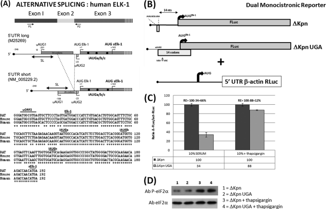
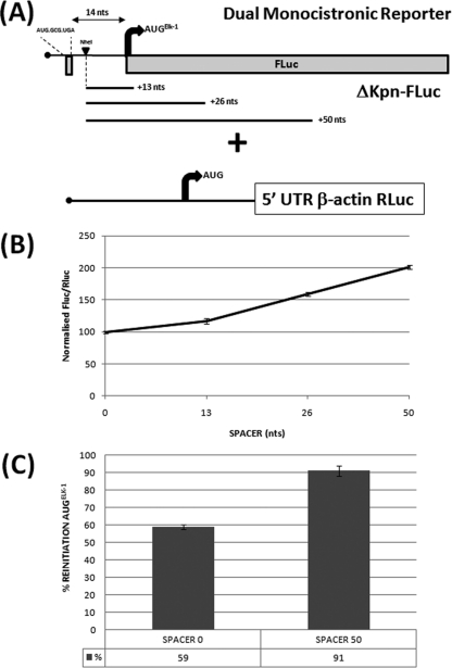
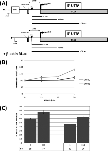
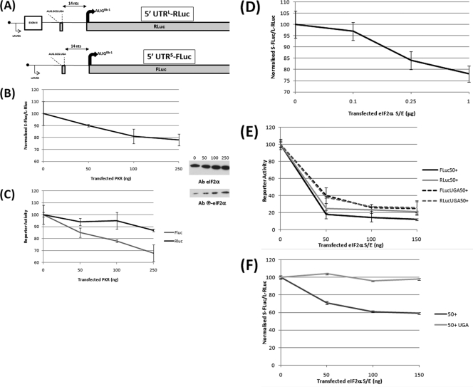
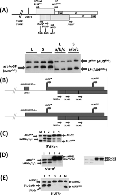
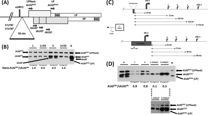
The 5' untranslated region (UTR) plays a central role in the regulation of mammalian translation initiation. Key components include RNA structure, upstream AUGs (uAUGs), upstream open reading frames (uORFs), and internal ribosome entry site elements that can interact to modulate the readout. We previously reported the characterization of two alternatively spliced 5' UTR isoforms of the human elk-1 gene. Both contain two uAUGs and a stable RNA stem-loop, but the long form (5' UTR(L)) was more repressive than the short form (5' UTR(S)) for initiation at the ELK-1 AUG. We now demonstrate that ELK-1 expression arises by a combination of leaky scanning and reinitiation, with the latter mediated by the small uORF2 conserved in both spliced isoforms. In HEK293T cells, a considerable fraction of ribosomes scans beyond the ELK-1 AUG in a reinitiation mode. These are sequestered by a series of out-of-frame AUG codons that serve to prevent access to a second in-frame AUG start site used to express short ELK-1 (sELK-1), an N-terminally truncated form of ELK-1 that has been observed only in neuronal cells. We present evidence that all these events are fine-tuned by the nature of the 5' UTR and the activity of the α subunit of eukaryotic initiation factor 2 and provide insights into the neuronal specificity of sELK-1 expression.


Figures

Fig 1
Fig 1
Schematic representation of the elk-1 gene. (A) (Top) The first three exons, with P1 and P2 indicating the primers used to characterize the alternatively spliced exon 2. Also depicted is the ORF organization and positioning of the AUG codons upstream of the AUGsELK-1 in the alternatively spliced transcripts. Note that in the 5′ UTRS, uAUG1 and uAUG2 are in the same ORF. SL refers to a stable stem-loop structure in which the uAUG1 is embedded. (Bottom) Alignment of the human, rat, and mouse sequences between the uORF2 and AUGsELK-1. (B) ΔKpn-FLuc and β-actin RLuc monocistronic reporter constructs. The box below the line depicts the small uORF2 with the sequence indicated. The impact of the UGA/C mutation in uORF2 on the reading frame organization relative to the AUGELK-1 is also depicted. (C) HEK293T cells were cotransfected with each ΔKpn-FLuc (WT and UGA/C) plasmid construct and the β-actin–RLuc control (referred to as the normalization plasmid). At 4 h posttransfection, medium was removed and replaced with DMEM containing 10% fetal calf serum with and without thapsigargin (300 nM). Cells were harvested after 24 h, and reporter activities were measured. Normalization of reporter activities was performed as outlined in Materials and Methods. The normalized FLuc/RLuc ratios are depicted graphically, with the WT value set at 100 for each condition tested. The value (relative to the WT) obtained in the UGA/C background represents that fraction of AUGELK-1 initiation events arising due to leaky scanning and is indicated below the graph (the difference between the WT and UGA/C values reflects the loss of reinitiation-mediated events). Reinitiation (RE) frequency at the AUGELK-1 is depicted above the data. (D) Immunoblot with eIF2α- and phospho-eIF2α-specific antibodies (Ab) from extracts tested as described above. All transfections were performed in triplicate, and the bars indicate the SEM.
Fig 2
Fig 2
Impact of spacer sequences downstream of uORF2 on initiation events at the AUGELK-1. (A) Schematic representation of the ΔKpn-FLuc/β-actin–RLuc reporters. The NheI indicates a unique restriction site introduced just downstream of the uORF2. The spacer elements of 13, 26, and 50 nt were inserted at this site. The 14 nt is the normal distance between the uORF2 and AUGELK-1. (B) Constructs were cotransfected into HEK293T cells with the β-actin–RLuc control. Normalization of reporter activities was performed as outlined in Materials and Methods. For the 5′ UTR spacer series, the SPACER 0 (no insert) value was set at 100. (C) Reinitiation frequency was estimated for SPACER 0 and SPACER 50 by comparing the normalized FLuc/RLuc ratios for the WT and UGA/C constructs, as outlined in the legend to Fig. 1. The mean percent reinitiation frequency is indicated below each column. All transfections were performed in triplicate, and the bars indicate the SEM. In panel C, P = 0.001.
Fig 3
Fig 3
Analysis of the uORF2 in the context of the alternative elk-1 5′ UTRs. (A) Schematic representation of the 5′ UTRL-FLuc and 5′ UTRS-FLuc reporters cotransfected into HEK293T cells with the β-actin–RLuc normalization control. The NheI site and spacer insertions are also indicated. The alternatively spliced exon 2 is indicated by a box. (B) The effect of the spacer sequences was assayed as outlined in the legend to Fig. 2B. (C). Reinitiation frequency was estimated for SPACER 0 and SPACER 50 by comparing the normalized FLuc/RLuc ratios for the WT and UGA/C constructs as outlined in the legend to Fig. 1. The mean percent reinitiation frequency is indicated below each column. All transfections were performed in triplicate, and the bars indicate the SEM. The P values for the S/S50 and L/L50 assays depicted in panel C were 0.02 and 0.003, respectively.
Fig 4
Fig 4
The 5′ UTRL and 5′ UTRS respond differently to changes in eIF2α activity. (A) Schematic representation of the Renilla (RLuc) and firefly (FLuc) reporter constructs. (B) HEK293T cells were cotransfected with the 5′ UTRL-RLuc and 5′ UTRS-FLuc constructs in the presence of increasing amounts of a plasmid expressing the PKR kinase. Normalized FLuc/RLuc activities were plotted against the amount of PKR plasmid transfected, with 0 ng being set at 100. (C) Values for each reporter, with 0 ng being set at 100. The inset on the right demonstrates that the PKR dose produced a progressive increase in the levels of phospho-eIF2α, as determined by immunoblotting. (D) The dual-reporter assay was performed in the presence of increasing amounts of a plasmid clone expressing eIF2αS/E. The FLuc/RLuc-normalized value for 0 μg eIF2αS/E was set at 100. (E and F) The eIF2αS/E dosage experiment was performed with 5′ UTRL-RLuc and 5′ UTRS-FLuc reporters carrying the SPACER 50 insertion with or without the UGA/C mutation of the uORF2. (E) Values for each reporter; (F) normalized values. All transfections were performed in triplicate, and the bars indicate the SEM.
Fig 5
Fig 5
The iAUGs serve to limit ribosomal access to the AUGsELK-1. (A) (Top) LP/SP reporter system that was adapted to monitor initiation events at the AUGELK-1, AUGsELK-1, and iAUG start codons. The position of the HA epitope in each ORF is indicated, as are the mutations introduced to remove the iAUG codons. The bars above and below the line indicate that LP and SP are in different ORFs. (Bottom) Immunoblot performed with the anti-HA antibody. HEK293T cells were transfected with the 5′ UTRL-LPNext (L), 5′ UTRS-LPNext (S), and the same 5′ UTRs carrying the iAUG → AGG changes (La/b/c and Sa/b/c). Transfections were performed in duplicate. (B) Schematic representation of the single nucleotide insertion within the uORF2 that fuses this ORF to the ORFs of AUGELK-1 and AUGsELK-1 (the LP ORF in the reporter). (C to E) Immunoblots performed with the anti-HA antibody for the three constructs tested. Lanes 1 and 2, WT constructs; lanes 3 and 4, iAUGa/b/c → AGG mutations. All clones were transfected in duplicate. The inset in panel D is a shorter exposure that demonstrates initiation products from both uAUG1 and uAUG2 (which are in the same ORF [Fig. 1A]). Lane M, a marker (the Sa/b/c construct depicted in panel A) for initiation products from the AUGELK-1 (LPNext) and AUGsELK-1 (LP).
Fig 6
Fig 6
The LP/SP reporter system demonstrates differences in the behavior of the 5′ UTRs. (A) Schematic representation of the SPACER 50 insertion in the context of 5′ UTRL-LPNext (L) and 5′ UTRS-LPNext (S). (B) The SPACER 0 (WT, represented by L and S) and SPACER 50 (L+50 and S+50) constructs were expressed in HEK293T cells. Proteins were visualized by immunoblotting with an anti-HA antibody. Lane Δ, a cell extract prepared from ΔKpn-LPNext-transfected cells that provides a marker for protein products arising from all the AUG codons depicted in panel A. Transfections were performed in duplicate. The protein bands in each individual transfection arising from the AUGELK-1 and iAUGa/b/c were quantified. The value of their ratio in each transfection is indicated below the gel (averaged from the duplicate samples). (C) Schematic representation of the impact of the uORF2 UGA/C mutation in the 5′ UTRL-LPNext and 5′ UTRS-LPNext reporter system. The sequence of the uORF2 is indicated, as is the distance between the stop codon of this ORF and the downstream AUG codons (arrows). (D) HEK293T cells were transfected with ΔKpn-LPNext (lanes Δ), 5′ UTRL-LPNext (lanes L), 5′ UTRS-LPNext (lanes S), and the long and short 5′ UTRs carrying the UGA/C mutation (lanes L-UGA/C and S-UGA/C). Protein products were detected by immunoblotting with the anti-HA antibody. Lane M, the same marker for LPNext and LP as outlined in the legend to Fig. 5. The AUGELK-1/iAUGa/b/c levels within each duplicate transfection were determined, and the averaged value is indicated below the gel. (Lower inset) An overexposure of the L-UGA/C and S-UGA/C lanes demonstrating the presence of a protein product arising from the AUGELK-1.
Fig 7
Fig 7
Expression from the AUGsELK-1 can be observed in HEK293T cells upon dosing of eIF2αS/E. The ΔKpn-LPNext (A), 5′ UTRL-LPNext (B), and 5′ UTRS-LPNext (C) constructs were transfected in the presence of increasing amounts (indicated in ng above each blot) of a plasmid expressing the negative eIF2αS/E. Each transfection was performed in duplicate. Proteins were visualized by immunoblotting with an anti-HA antibody. Lane C, a mock-transfected control; lane M, a marker for LPNext (AUGELK-1) and LP (AUGsELK-1). (D) Negative effect of the eIF2αS/E dosage on an LP-expressing plasmid with a short 5′ UTR. The LP protein was detected by immunoblotting with an anti-HA antibody. The steady-state levels of eIF2α were followed with an anti-eIF2α antibody.

Similar articles

Cited by

References

    1. Andreev DE, et al. 2009. Differential contribution of the m7G-cap to the 5′ end-dependent translation initiation of mammalian mRNAs. Nucleic Acids Res. 37:6135–6147 - PMC - PubMed
    1. Araud T, Genolet R, Jaquier-Gubler P, Curran J. 2007. Alternatively spliced isoforms of the human elk-1 mRNA within the 5′ UTR: implications for ELK-1 expression. Nucleic Acids Res. 35:4649–4663 - PMC - PubMed
    1. Calkhoven CF, Muller C, Leutz A. 2000. Translational control of C/EBPalpha and C/EBPbeta isoform expression. Genes Dev. 14:1920–1932 - PMC - PubMed
    1. Churbanov A, Rogozin IB, Babenko VN, Ali H, Koonin EV. 2005. Evolutionary conservation suggests a regulatory function of AUG triplets in 5′-UTRs of eukaryotic genes. Nucleic Acids Res. 33:5512–5520 - PMC - PubMed
    1. Costa-Mattioli M, et al. 2007. eIF2alpha phosphorylation bidirectionally regulates the switch from short- to long-term synaptic plasticity and memory. Cell 129:195–206 - PMC - PubMed

Publication types

Substances

LinkOut - more resources