. 2012 Mar 6;109(10):3826-31.
doi: 10.1073/pnas.1115201109. Epub 2012 Feb 21.

Cyclin-dependent kinase 1 (Cdk1) is essential for cell division and suppression of DNA re-replication but not for liver regeneration

Affiliations

Cyclin-dependent kinase 1 (Cdk1) is essential for cell division and suppression of DNA re-replication but not for liver regeneration

M Kasim Diril et al. Proc Natl Acad Sci U S A. .

Abstract

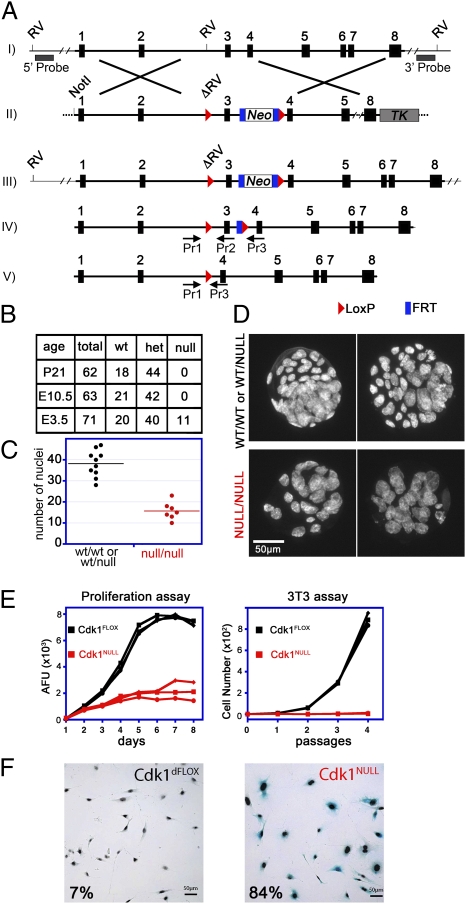
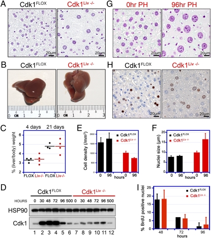
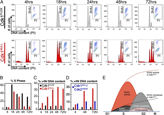
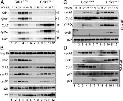
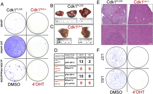
Cyclin-dependent kinase 1 (Cdk1) is an archetypical kinase and a central regulator that drives cells through G2 phase and mitosis. Knockouts of Cdk2, Cdk3, Cdk4, or Cdk6 have resulted in viable mice, but the in vivo functions of Cdk1 have not been fully explored in mammals. Here we have generated a conditional-knockout mouse model to study the functions of Cdk1 in vivo. Ablation of Cdk1 leads to arrest of embryonic development around the blastocyst stage. Interestingly, liver-specific deletion of Cdk1 is well tolerated, and liver regeneration after partial hepatectomy is not impaired, indicating that regeneration can be driven by cell growth without cell division. The loss of Cdk1 does not affect S phase progression but results in DNA re-replication because of an increase in Cdk2/cyclin A2 activity. Unlike other Cdks, loss of Cdk1 in the liver confers complete resistance against tumorigenesis induced by activated Ras and silencing of p53.


Conflict of interest statement

The authors declare no conflict of interest.

Figures

Fig. 1.
Fig. 1.
Generation and analysis of Cdk1 conditional knockout mice and cells. (A) The Cdk1 genomic locus (I) was modified in ES cells with the targeting vector (II) shown. An FRT-flanked neomycin-selection cassette was introduced along with LoxP recombination sites (red triangles) on both sides of exon 3, which generated a mutant Cdk1 locus (III). For Southern blot analysis, 5′ and 3′ probes located outside of the targeting vector were used, after an EcoRV (RV) digest, resulting in a 31,206-bp (recombinant) and a 9,110-bp (5′) or 20,300-bp (3′) fragment for wild type (Fig. S1A). Upon expression of FLP recombinase, the neomycin cassette was removed, and only the LoxP sites remained in the locus (IV, Cdk1FLOX). The levels of Cdk1 protein expression were similar in Cdk1FLOX and Cdk1WT mice (Fig. S1B). After Cre recombinase expression, exon 3 was excised (V), which resulted in deletion of Cdk1 and a frame shift. PCR genotyping primers are indicated (Pr1, -2, and -3), and the sequences can be found in Table S1. (B) To generate Cdk1 knockouts, Cdk1FLOX mice were crossed with β-actin–Cre mice expressing Cre recombinase ubiquitously in all tissues, including germ line. The resulting Cdk1WT/NULL mice were interbred, and the offspring was analyzed at weaning (P21), midgestation (E10.5), or blastocyst (E3.5) stage. (C and D) Cdk1NULL/NULL blastocysts were visualized by Hoechst staining of their nuclei followed by fluorescence microscopy. Comparison of Cdk1-deficient blastocysts with heterozygous or wild-type littermates indicated a reduced number of cells (C); however, their nuclei were larger in size (D). (E and F) Three independent MEF lines (Fig. S1 CH) were treated with 4-OHT to induce Cdk1 knockout, and their proliferative potential was monitored by 3T3 and alamarBlue proliferation assays for several passages or days, respectively (E). Deletion of Cdk1 resulted in a rapid arrest of cellular proliferation and premature onset of cellular senescence that was detected by senescence-associated β-gal staining (F). Therefore, cells lacking Cdk1 cannot proliferate but instead enter a senescent state and survive in culture medium.
Fig. 2.
Fig. 2.
Liver-specific knockout of Cdk1. (A) Liver sections from Cdk1Liv−/− and control mice were stained with Feulgen to visualize their nuclei. Even though the overall liver size was similar, Cdk1-knockout livers contained fewer hepatocytes with enlarged nuclei. (B and C) To analyze the regenerative potential and S phase entry in Cdk1-knockout livers, 70% of the liver mass was removed by PH. Animals were euthanized 4 or 21 d later, and the ratio of liver to body weight was determined. Each dot in the chart represents an individual PH experiment, and mean values are depicted by the black or red lines. Cdk1Liv−/− livers regenerated the lost mass within 3 wk, comparable to controls. (D) To confirm Cdk1 knockout, Western blots with Cdk1 antibodies were performed at different times after PH in control and Cdk1Liv−/− liver extracts. Cdk1 expression detected in knockout livers is due to liver cells other than hepatocytes. (E–G) Cdk1Liv−/− display a decreased hepatocyte density compared with controls and the difference is exacerbated by 96 h after PH (E). Cdk1Liv−/− hepatocytes have enlarged nuclei that become even bigger after PH (F and G). (H and I) Regenerating livers were pulse-labeled with BrdU at 48 (H), 72, and 96 h after PH. BrdU incorporation was detected by immunohistochemical staining of liver sections (H) and quantified with a custom developed image analysis software (I). Data were obtained from n = 3 animals in E, F, and I and are represented as mean ± SD. Black and red bars indicate Cdk1FLOX and Cdk1Liv−/− livers, respectively.
Fig. 3.
Fig. 3.
Cdk1 is redundant for S phase progression, but its loss results in endoreduplication. (A) Cdk1FLOX MEFs were synchronized in the G0/G1 phase by serum starvation for 72 h and simultaneously treated with 4-OHT to induce knockout of Cdk1. They were then released into S phase by addition of serum, pulse-labeled with BrdU, and analyzed by FACS to determine the percentage of cells in S, G1, and G2 phases. Histograms depict the distribution of DNA content as detected by propidium iodide (PI) staining. Insets show the distribution of BrdU incorporation (BrdU) versus DNA content (PI) within the same population. Three independent experiments were performed with three different clones. (B) Quantitative analysis of cells in S phases of the cell cycle are shown. Although Cdk1-knockout MEFs seem to have a slightly reduced ratio in S phase at 18 and 24 h, this is due to the accumulation of G2-arrested cells during the serum starvation period, which cannot enter S phase as efficiently as G1 cells. Data are represented as mean ± SD. (C) Deletion of Cdk1 resulted in endoreduplication and accumulation of cells with a DNA content greater than 4N. (D) However, simultaneous removal of Cdk2 with shRNAs partially rescued the endoreduplication phenotype. See also Fig. S3. (E) A model demonstrating how Cdk1 manages to reduce Cdk2 activity at the end of S phase and prevents DNA re-replication. According to our model, kinase activity of Cdk2/cyclin A2 complexes is higher than that of Cdk1/cyclin A2 complexes. Cyclin A2-associated kinase activity peaks in S phase. However, increased levels of Cdk1 protein during late S to early G2 phase results in fewer cyclin A2 molecules available for binding to Cdk2. As a result, Cdk1 quenches cyclin A2-associated kinase activity. In Cdk1-deficient cells (represented by dashed lines), cyclin A2 molecules are forming complexes only with Cdk2. Therefore, Cdk2/cyclin A2 kinase activity cannot be suppressed and persists throughout G2 phase.
Fig. 4.
Fig. 4.
Increased Cdk2 kinase activity in Cdk1-deficient MEFs. (A) To analyze the kinase activities associated with Cdks and cyclins, protein extracts were prepared from cells in Fig. 3 at different time points and subjected to immunoprecipitation with the indicated antibodies followed by in vitro kinase assays using radiolabeled ATP and histone H1 as substrates. Cdk2 and cyclin A2-associated kinase activities were significantly increased in Cdk1-deficient cells. (B) Samples from A were subjected to SDS/PAGE and Western blotting with the indicated antibodies. (C and D) To analyze the Cdk2/cyclinA2 complexes, protein extracts were immunoprecipitated with antibodies against Cdk2 (C) or cyclin A2 (D) followed by Western blotting with the indicated antibodies. See also Figs. S4S6 and S8.
Fig. 5.
Fig. 5.
Oncogenic transformation or tumorigenesis is not possible in the absence of Cdk1. (A) Cdk1FLOX MEFs were immortalized or transformed with p53 shRNA or activated Ras/p53DN, respectively. Colony-formation assays after induction of the cells with 4-OHT indicate that Cdk1-deficient cells cannot be immortalized or transformed. (B–E) Liver tumors were induced by tail-vein injection of activated Ras (B), but no tumors were detected in Cdk1Liv−/− livers (C). Quantitative analysis of liver tumors in mice of the indicated genotypes (D) is shown. Histological sections of the liver were stained with hematoxylin and eosin (E) to demonstrate normal morphology in the Cdk1Liv−/− (Right) and many tumors in control livers (Left). (F) Tumors from two different Cdk1FLOX/FLOX mice (LI37 and LI53) were isolated, dissociated, and grown in culture. After introduction of CreERT2 and treatment with DMSO (control) or 4-OHT, cells were subjected to a colony-formation assay followed by Giemsa staining.

Similar articles

Cited by

References

    1. Morgan DO. The Cell Cycle: Principles of Control. London: New Science Press; 2007.
    1. Malumbres M, Barbacid M. Cell cycle, CDKs and cancer: A changing paradigm. Nat Rev Cancer. 2009;9:153–166. - PubMed
    1. Satyanarayana A, Kaldis P. Mammalian cell-cycle regulation: Several Cdks, numerous cyclins and diverse compensatory mechanisms. Oncogene. 2009;28:2925–2939. - PubMed
    1. Lohka MJ, Hayes MK, Maller JL. Purification of maturation-promoting factor, an intracellular regulator of early mitotic events. Proc Natl Acad Sci USA. 1988;85:3009–3013. - PMC - PubMed
    1. Nurse P, Thuriaux P. Regulatory genes controlling mitosis in the fission yeast Schizosaccharomyces pombe. Genetics. 1980;96:627–637. - PMC - PubMed

Publication types