. 2012 Mar 6;109(10):3838-43.
doi: 10.1073/pnas.1006619109. Epub 2012 Feb 21.

Full regeneration of the tribasal Polypterus fin

Affiliations

Full regeneration of the tribasal Polypterus fin

Rodrigo Cuervo et al. Proc Natl Acad Sci U S A. .

Abstract

Full limb regeneration is a property that seems to be restricted to urodele amphibians. Here we found that Polypterus, the most basal living ray-finned fish, regenerates its pectoral lobed fins with a remarkable accuracy. Pectoral Polypterus fins are complex, formed by a well-organized endoskeleton to which the exoskeleton rays are connected. Regeneration initiates with the formation of a blastema similar to that observed in regenerating amphibian limbs. Retinoic acid induces dose-dependent phenotypes ranging from inhibition of regeneration to apparent anterior-posterior duplications. As in all developing tetrapod limbs and regenerating amphibian blastema, Sonic hedgehog is expressed in the posterior mesenchyme during fin regeneration. Hedgehog signaling plays a role in the regeneration and patterning processes: an increase or reduction of fin bony elements results when this signaling is activated or disrupted, respectively. The tail fin also regenerates but, in contrast with pectoral fins, regeneration can resume after release from the arrest caused by hedgehog inhibition. A comparative analysis of fin phenotypes obtained after retinoic acid treatment or altering the hedgehog signaling levels during regeneration allowed us to assign a limb tetrapod equivalent segment to Polypterus fin skeletal structures, thus providing clues to the origin of the autopod. We propose that appendage regeneration was a common property of vertebrates during the fin to limb transition.


Conflict of interest statement

The authors declare no conflict of interest.

Figures
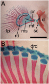
Fig. 1.
Fig. 1.
Anatomy of the Polypterus fin. (A) Frontal view of alcian blue- and alizarin red-stained right fin and pectoral girdle, indicating the main fin skeletal components: cleithrum (cl), lepidotrichia (lp), mesopterygium (ms), metapterygium (mt), propterygium (pr), radials (rd), and scapulocoracoid (sc). (Scale bar, 5 mm.) Note the small bone element (*) that articulates to each of the large bone elements metapterygium and propterygium. Segments that may be equivalent to the autopod and zeugopod of tetrapods are also indicated (autopodial- and zeugopodial-like; Discussion). (B) Close-up of A to visualize the distal radials (drd) attached to each radial bone element. Posterior is to the bottom in A and to the left in B.
Fig. 2.
Fig. 2.
Pectoral fin regeneration in P. ornatipinnis and fin endoskeleton regeneration in P. senegalus. (A) External appearance of the same fin during the regeneration process at sequential days after amputation. “d0” shows a fin before amputation. Anterior is to the right, and dorsal to the top. (Scale bar, 8 mm.) (B) Alcian blue- and alizarin red-stained regenerates at different days after amputation (see also Fig. S1). Anterior is to the left. (Scale bar, 2.5 mm.) (C) External appearance of blastema and apical fold in a 4-day regenerate. (D) Toluidine blue-stained longitudinal sections of regenerates at different days after amputation. Lower: Areas within the squares (Upper), shown at higher magnification. Arrowhead in the 4d high-magnification picture indicates the apical fold, a structure formed by basal stratum cells. Ventral is to the right. (Scale bars, 400 μm in Upper, 40 μm in Lower.) bs, basal stratum; cc, cartilage condensation; cp, cartilaginous plate; eg, external gill.
Fig. 3.
Fig. 3.
Fin regenerates treated with RA. (A) Ventral view of right fin regenerates treated with 100 μg/g RA at 3, 6, and 9 dpa, as described in Materials and Methods. (B) Ventral view of a right fin treated with 75 μg/g RA at 3 dpa, showing a phenotype resembling AP duplications; arrowhead indicates a bend near an incipient second point of duplication. Right: Enlarged area shows a different view of the putative second duplication; note the small lepidotrichia (arrowheads) shared by the adjacent fin lobe and flanking small lobule. (C) Ventral view of a left fin treated with 75 μg/g RA at 3 dpa showing what appears to be the emergence of a large cartilage, possibly corresponding to a duplicated propterigium or metapterigium; arrowheads indicate lepidotrichia emerging from the new large cartilage. Right: Enlarged area shows a dorsal view of the same fin. Arrows indicate the point of main duplication. All fins shown were dissected and stained with alcian blue and alizarin red 35 d after RA treatment.
Fig. 4.
Fig. 4.
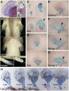
Fin regenerates treated with cyclopamine and Shh agonist. (A) Frontal view of a 10 dpa right fin regenerate hybridized with an Shh probe (same position as Fig. 1A). Arrows point to the cleithrum bone and scapula. (B) Ventral and dorsal view of control (Top and Bottom Left, respectively) and cyclopamine-treated (Middle and Bottom Right, respectively) pectoral fins after 35 d of regeneration. (C) Skeletal staining of early regenerates treated with 2 μg/mL cyclopamine (Cyc). Right: Magnifications of fins shown at Left. Arrowhead marks the limit of the anterior (to the right) region affected. (D) Skeletal staining of late regenerates treated with 1 μg/mL cyclopamine. (E) Skeletal staining of an early regenerate treated with DMSO (Control). (F) Skeletal staining of regenerates treated with 100 nM Hh-Ag1.3. (G) Skeletal staining of a regenerate treated with 200 nM Hh-Ag1.3. Arrowhead marks the AP limit at which the posterior region appears more affected. (H) Skeletal staining of left fin regenerates and pectoral girdles of P. ornatipinnis after treatment with 2 μg/mL cyclopmine at 8, 10, 12, and 14 dpa and then left regenerating for 3 wk; control is an untreated fin regenerated for 42 d. Note the sequential reduction of the mesopterygial plate and lepidotrichia, with minor disturbances of propterygium and metapterygium bones. Materials and Methods provides details about treatment protocols. a, anterior; cl, cleithrum; d, dorsal; p, posterior; sc, scapulocoracoid; v, ventral.
Fig. 5.
Fig. 5.
Effects of altering Shh signaling on regeneration of fin tail lepidotrichia. (A) Alizarin red staining of control (DMSO-treated) tail fin. (B) Tail fin treated with 2 μg/mL cyclopamine. (C) Tail fin treated with 100 μM of Shh agonist. (D) Regeneration of tail fin before and 3 wk after cyclopamine withdrawal. Fin pictures were taken at 3 wk after amputation, except for the picture in D. Cyclopamine impairs the regenerative process (A and B), but it is resumed after cyclopamine withdrawal (D). The Hh-Ag3.1 agonist did not affect the number, but the morphology of the regenerating tail fin lepidotrichia showed a zigzag pattern (C).

Similar articles

Cited by

References

    1. Coates MI. The origin of vertebrate limbs. Dev Suppl. 1994:169–180. - PubMed
    1. Bartsch P, Gemballa S, Piotrowski T. The embryonic and larval development of Polypterus senegalus Cuvier 1829: Its staging by external and skeletal features, behaviour and undulatory locomotion. Acta Zoologica. 1997;78:309–328.
    1. Clack JA. Earliest known tetrapod braincase and the evolution of the stapes and fenestra ovalis. Nature. 1994;369:392–394.
    1. Coates MI, Clack JA. Fish-like gills and breathing in the earliest known tetrapod. Nature. 1991;352:234–236.
    1. Budgett JS. On the structure of the larval Polypterus. T Zool Soc London. 1902;16:315–338.

Publication types

Associated data