

Rat brains also have a default mode network
- PMID: 22355129
- PMCID: PMC3309754
- DOI: 10.1073/pnas.1200506109
Rat brains also have a default mode network
Abstract
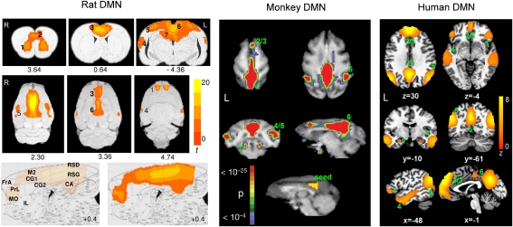
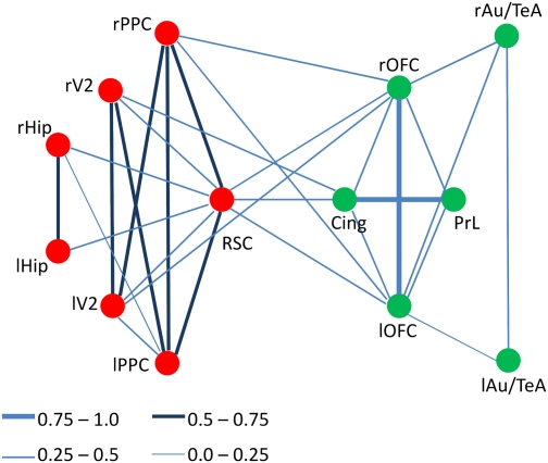
The default mode network (DMN) in humans has been suggested to support a variety of cognitive functions and has been implicated in an array of neuropsychological disorders. However, its function(s) remains poorly understood. We show that rats possess a DMN that is broadly similar to the DMNs of nonhuman primates and humans. Our data suggest that, despite the distinct evolutionary paths between rodent and primate brain, a well-organized, intrinsically coherent DMN appears to be a fundamental feature in the mammalian brain whose primary functions might be to integrate multimodal sensory and affective information to guide behavior in anticipation of changing environmental contingencies.
Conflict of interest statement
The authors declare no conflict of interest.
Figures
Similar articles
-
Constituents and functional implications of the rat default mode network.Proc Natl Acad Sci U S A. 2016 Aug 2;113(31):E4541-7. doi: 10.1073/pnas.1601485113. Epub 2016 Jul 20. Proc Natl Acad Sci U S A. 2016. PMID: 27439860 Free PMC article.
-
Test-retest reproducibility of the default-mode network in healthy individuals.Hum Brain Mapp. 2010 Feb;31(2):237-46. doi: 10.1002/hbm.20860. Hum Brain Mapp. 2010. PMID: 19621371 Free PMC article.
-
Does the default-mode functional connectivity of the brain correlate with working-memory performances?Arch Ital Biol. 2009 Mar;147(1-2):11-20. Arch Ital Biol. 2009. PMID: 19678593
-
Default mode network activity and connectivity in psychopathology.Annu Rev Clin Psychol. 2012;8:49-76. doi: 10.1146/annurev-clinpsy-032511-143049. Epub 2012 Jan 6. Annu Rev Clin Psychol. 2012. PMID: 22224834 Review.
-
Default mode network activity in bipolar disorder.Epidemiol Psychiatr Sci. 2020 Sep 8;29:e166. doi: 10.1017/S2045796020000803. Epidemiol Psychiatr Sci. 2020. PMID: 32895076 Free PMC article. Review.
Cited by
-
Human hippocampal connectivity is stronger in olfaction than other sensory systems.Prog Neurobiol. 2021 Jun;201:102027. doi: 10.1016/j.pneurobio.2021.102027. Epub 2021 Feb 25. Prog Neurobiol. 2021. PMID: 33640412 Free PMC article.
-
The restless brain: how intrinsic activity organizes brain function.Philos Trans R Soc Lond B Biol Sci. 2015 May 19;370(1668):20140172. doi: 10.1098/rstb.2014.0172. Philos Trans R Soc Lond B Biol Sci. 2015. PMID: 25823869 Free PMC article. Review.
-
Preclinical Magnetic Resonance Imaging and Spectroscopy Studies of Memory, Aging, and Cognitive Decline.Front Aging Neurosci. 2016 Jun 29;8:158. doi: 10.3389/fnagi.2016.00158. eCollection 2016. Front Aging Neurosci. 2016. PMID: 27468264 Free PMC article. Review.
-
The effects of alcohol on the nonhuman primate brain: a network science approach to neuroimaging.Alcohol Clin Exp Res. 2013 Nov;37(11):1891-900. doi: 10.1111/acer.12181. Epub 2013 Jul 26. Alcohol Clin Exp Res. 2013. PMID: 23905720 Free PMC article.
-
Resting state network topology of the ferret brain.Neuroimage. 2016 Dec;143:70-81. doi: 10.1016/j.neuroimage.2016.09.003. Epub 2016 Sep 2. Neuroimage. 2016. PMID: 27596024 Free PMC article.
References
-
- Buckner RL, Andrews-Hanna JR, Schacter DL. The brain's default network: Anatomy, function, and relevance to disease. Ann N Y Acad Sci. 2008;1124:1–38. - PubMed
-
- Binder JR, et al. Conceptual processing during the conscious resting state. A functional MRI study. J Cogn Neurosci. 1999;11:80–95. - PubMed
-
- Vincent JL, et al. Intrinsic functional architecture in the anaesthetized monkey brain. Nature. 2007;447:83–86. - PubMed
Publication types
MeSH terms
Grants and funding
LinkOut - more resources
Full Text Sources
