. 2012 Mar 6;109(10):3784-9.
doi: 10.1073/pnas.1117768109. Epub 2012 Feb 21.

The distribution of ligand-binding pockets around protein-protein interfaces suggests a general mechanism for pocket formation

Affiliations

The distribution of ligand-binding pockets around protein-protein interfaces suggests a general mechanism for pocket formation

Mu Gao et al. Proc Natl Acad Sci U S A. .

Abstract

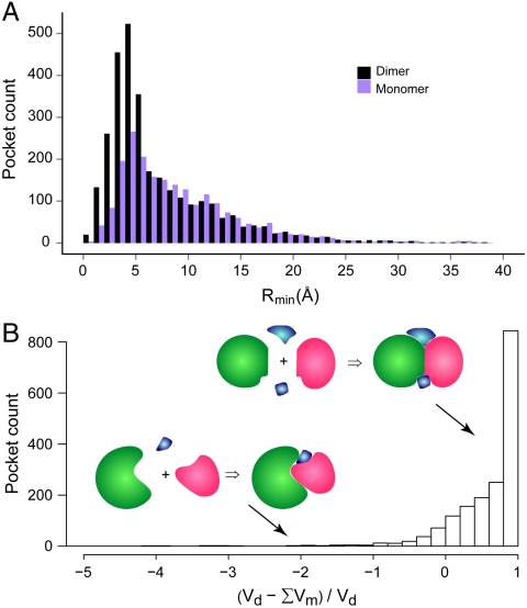
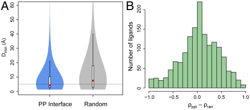
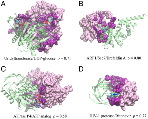
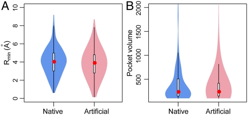
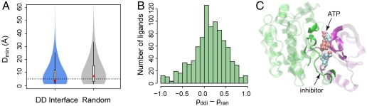
Protein-protein and protein-ligand interactions are ubiquitous in a biological cell. Here, we report a comprehensive study of the distribution of protein-ligand interaction sites, namely ligand-binding pockets, around protein-protein interfaces where protein-protein interactions occur. We inspected a representative set of 1,611 representative protein-protein complexes and identified pockets with a potential for binding small molecule ligands. The majority of these pockets are within a 6 Å distance from protein interfaces. Accordingly, in about half of ligand-bound protein-protein complexes, amino acids from both sides of a protein interface are involved in direct contacts with at least one ligand. Statistically, ligands are closer to a protein-protein interface than a random surface patch of the same solvent accessible surface area. Similar results are obtained in an analysis of the ligand distribution around domain-domain interfaces of 1,416 nonredundant, two-domain protein structures. Furthermore, comparable sized pockets as observed in experimental structures are present in artificially generated protein complexes, suggesting that the prominent appearance of pockets around protein interfaces is mainly a structural consequence of protein packing and thus, is an intrinsic geometric feature of protein structure. Nature may take advantage of such a structural feature by selecting and further optimizing for biological function. We propose that packing nearby protein-protein or domain-domain interfaces is a major route to the formation of ligand-binding pockets.


Conflict of interest statement

The authors declare no conflict of interest.

Figures

Fig. 1.
Fig. 1.
Distribution of pockets from protein-protein interfaces. Pockets are calculated using dimeric complex structures (denoted as “dimer”) and individual monomeric structures, respectively. (A) Histograms of pockets versus the distance from the protein interface. The width of the distance bins is 1 Å; few cases with extreme values > 40  are not shown. Definition of Rmin is given in the text. (B) Statistics of volume changes for interfacial pockets with a Rmin < 6 . The volumes of a pocket found in dimers and separated monomers are denoted as Vd and Vm, respectively. The summation is over all monomer pockets associated with each dimer pocket (see text). Insets are diagrams that depict ligand-binding pockets formed upon protein-protein complexation. Ligands are colored in blue and the two proteins are colored in green and red.
Fig. 2.
Fig. 2.
The distribution of ligands from protein-protein interfaces. (A) Violin plot of the minimal distance between ligand and protein interface/random surface patch. The plot is derived from a boxplot by scaling the width of the box, such that the area is proportional to the number of structures observed. A dotted horizontal line is located at a Dmin of 5 Å. The white bars range from 25th to 75th percentile; and whiskers extend to a distance of up to 1.5 times the interquartile range. The red spheres represent the medians. The same violin plot schemes are employed in subsequent figures. (B) Histogram of the difference in the fraction of ligand contact surface area contributed by protein interface residues versus random surface residues. Only cases with nonzero values are shown.
Fig. 3.
Fig. 3.
Examples of ligands bound at protein-protein interfaces. Protein and ligand are (A) Uridyltransferase/UDP-glucose (PDB code: 1guq), (B) ARF1/Sec7/Brefeldin A (1re0), (C) ATPase P4/ATP analog (1w48), (D) HIV-1 protease/Ritonavir (1rl8). In each snapshot, one protein monomer is shown in a surface representation, where interfacial/noninterfacial residues are shown in dark/light purple colors, respectively; for clarity, the other protein monomer is shown in a green cartoon representation. The ligand is shown in a van der Waals representation using the following color code: carbon (cyan), nitrogen (blue), oxygen (red), sulfur (yellow), and phosphate (tan). Molecular images were created with VMD (42).
Fig. 4.
Fig. 4.
Pockets of native complexes versus pockets of artificial complexes. (A) Distribution of pockets in the neighborhood of protein interfaces. (B) Comparison of pocket volume defined as the number of grid points.
Fig. 5.
Fig. 5.
Ligand distributions around protein domain-domain interfaces. (A) The minimal distance from ligand to domain interface versus the distance to random surface patch. (B) The fraction of ligand contact surface area contributed by domain interface versus that of a random surface. (C) Example of a ligand bound to a protein domain interface. The N- and C-terminal domains of the protein kinase MEK1 are shown in purple and green cartoon representations, respectively. Two ligands cocrystallized are shown in a vdW representation. Protein residues contacting the ligands are displayed in solid colors, and other residues are dimmed for clarity.

Similar articles

Cited by

References

    1. Alberts B. Molecular biology of the cell. 5th Ed. New York: Garland Science; 2008.
    1. Berman HM, et al. The Protein Data Bank. Nucleic Acids Res. 2000;28:235–242. - PMC - PubMed
    1. Janin J, Bahadur RP, Chakrabarti P. Protein-protein interaction and quaternary structure. Q Rev Biophys. 2008;41:133–180. - PubMed
    1. Jones S, Thornton JM. Principles of protein-protein interactions. Proc Natl Acad Sci USA. 1996;93:13–20. - PMC - PubMed
    1. Keskin Z, Gursoy A, Ma B, Nussinov R. Principles of protein-protein interactions: What are the preferred ways for proteins to interact? Chem Rev. 2008;108:1225–1244. - PubMed

Publication types

LinkOut - more resources