. 2012 Mar 6;109(10):3754-9.
doi: 10.1073/pnas.1113384109. Epub 2012 Feb 21.

Chromosome segregation in Archaea mediated by a hybrid DNA partition machine

Affiliations

Chromosome segregation in Archaea mediated by a hybrid DNA partition machine

Anne K Kalliomaa-Sanford et al. Proc Natl Acad Sci U S A. .

Abstract

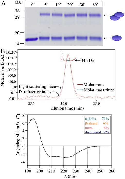
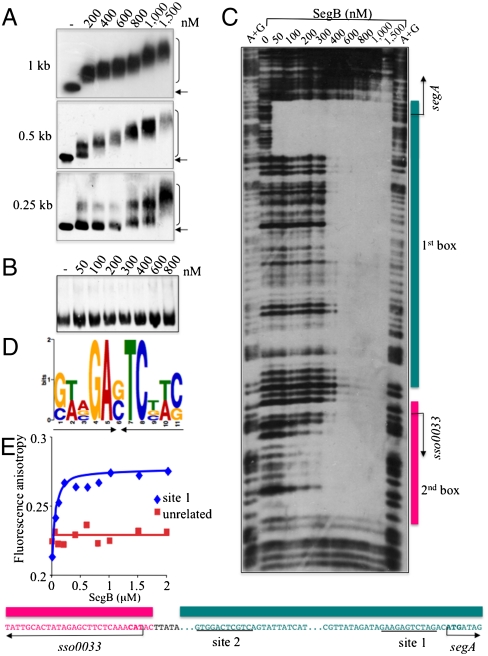
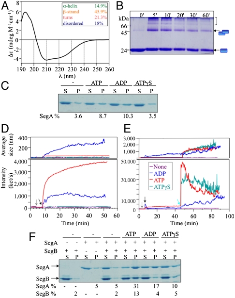
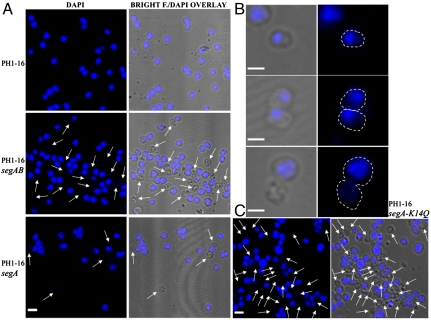
Eukarya and, more recently, some bacteria have been shown to rely on a cytoskeleton-based apparatus to drive chromosome segregation. In contrast, the factors and mechanisms underpinning this fundamental process are underexplored in archaea, the third domain of life. Here we establish that the archaeon Sulfolobus solfataricus harbors a hybrid segrosome consisting of two interacting proteins, SegA and SegB, that play a key role in genome segregation in this organism. SegA is an ortholog of bacterial, Walker-type ParA proteins, whereas SegB is an archaea-specific factor lacking sequence identity to either eukaryotic or bacterial proteins, but sharing homology with a cluster of uncharacterized factors conserved in both crenarchaea and euryarchaea, the two major archaeal sub-phyla. We show that SegA is an ATPase that polymerizes in vitro and that SegB is a site-specific DNA-binding protein contacting palindromic sequences located upstream of the segAB cassette. SegB interacts with SegA in the presence of nucleotides and dramatically affects its polymerization dynamics. Our data demonstrate that SegB strongly stimulates SegA polymerization, possibly by promoting SegA nucleation and accelerating polymer growth. Increased expression levels of segAB resulted in severe growth and chromosome segregation defects, including formation of anucleate cells, compact nucleoids confined to one half of the cell compartment and fragmented nucleoids. The overall picture emerging from our findings indicates that the SegAB complex fulfills a crucial function in chromosome segregation and is the prototype of a DNA partition machine widespread across archaea.


Conflict of interest statement

The authors declare no conflict of interest.

Figures

Fig. 1.
Fig. 1.
SegB is a dimeric, helical protein. (A) 15% SDS-gel showing the products of a time-course crosslinking experiment in which SegB (10 μM) was incubated with DMP (10 mM). (B) SEC-MALLS analysis of SegB. The protein (10 μM) was injected into a Superdex 200 HR10/30 column and the elution profile monitored by differential refractive index (dashed line) and Rayleigh light scattering (continuous line). The brown line at the top of the peak is the derived molar mass and the green tract is the portion used for the peak molar mass calculation. (C) CD spectrum of SegB (10 μM) recorded at 30 °C and calculated percentages of secondary structure elements.
Fig. 2.
Fig. 2.
SegB is a site-specific DNA-binding protein. (A) EMSAs in which biotinylated DNA fragments (1–5 nM) spanning the region upstream of segA were incubated with SegB in the presence of competitor polydIdC DNA (1 μg). Arrow: free DNA; bracket: SegB•DNA complexes. (B) EMSA control experiment including an unrelated DNA fragment. (C) Top, DNase I footprinting showing the regions protected by SegB; Bottom, diagram illustrating the SegB protection regions and corresponding DNA sequence. Site 1 and 2 are underlined. (D) Logo of the palindromic motif identified through MEME (20). (E) Fluorescence anisotropy assay measuring binding of SegB to a double-stranded [Cy3]-labeled oligonucleotide (23 bp) (5 nM) containing the motif (site 1) (blue line) and to an unrelated oligonucleotide (5 nM) (red line) in the presence of competitor polydIdC DNA (50 μg/mL).
Fig. 3.
Fig. 3.
SegA assembles into extensive polymers, whose growth and dynamics are affected by SegB. (A) CD spectrum of SegA (10 μM) recorded at 30 °C and percentages of secondary structure elements. (B) 15% SDS-gel showing a DMP crosslinking time-course of SegA (7 μM) in the presence of ATP (2 mM). The bracket at the top indicates higher oligomers. (C) Sedimentation assay in which SegA (18 μM) was incubated with and without nucleotides (4 mM) at 30 °C and then centrifuged. 100% of the pellet (P) and 20% of the supernatant (S) fractions were resolved on a 15% SDS-gel. (D) SegA polymerization followed by DLS. Bottom illustrates the increase in light scattering intensity, expressed as kct/s; Top shows the corresponding augmentation in polymer average size (nm). SegA (5 μM) was incubated at 30 °C with and without nucleotides (ADP/ATP/ATPγS) (1 mM). The arrows indicate the point at which MgCl2 (1 mM) (gray) and nucleotides (black) were added. (E) SegB promotes SegA polymerization. DLS kinetics showing the effect SegB (5 μM) in the presence and absence of nucleotides. The green arrow indicates the time of SegB addition. Note the substantial difference in vertical scale in panel D and E. (F) Sedimentation assay in which SegA (12 μM) and SegB (12 μM) were coincubated with and without nucleotides.
Fig. 4.
Fig. 4.
Increased gene dosage of segA and segB results in a high rate of anucleate cells and anomalous nucleoid morphology in S. solfataricus. Phase contrast and fluorescence microscopy of DAPI-stained cells expressing higher levels of segAB and segA (A) or segA-K14Q (C). The arrows point to anucleate cells. Bar = 2 μm. (B) Examples of aberrant chromosome segregation phenotypes observed for the strain with increased levels of SegAB. Bar = 1 μm.

Similar articles

Cited by

References

    1. Kotwaliwale C, Biggins S. Microtubule capture: a concerted effort. Cell. 2006;127:1105–1108. - PubMed
    1. Hayes F, Barillà D. The bacterial segrosome: a dynamic nucleoprotein machine for DNA trafficking and segregation. Nat Rev Microbiol. 2006;4:133–143. - PubMed
    1. Schumacher MA. Structural biology of plasmid partition: uncovering the molecular mechanisms of DNA segregation. Biochem J. 2008;412:1–18. - PubMed
    1. Barillà D, Rosenberg MF, Nobbmann U, Hayes F. Bacterial DNA segregation dynamics mediated by the polymerizing protein ParF. EMBO J. 2005;24:1453–1464. - PMC - PubMed
    1. Lim GE, Derman AI, Pogliano J. Bacterial DNA segregation by dynamic SopA polymers. Proc Natl Acad Sci USA. 2005;102:17658–17663. - PMC - PubMed

Publication types

MeSH terms

LinkOut - more resources