. 2012 Mar 20;109(12):E698-704.
doi: 10.1073/pnas.1201107109. Epub 2012 Feb 21.

Evidence for an intermediate conformational state of LacY

Affiliations

Evidence for an intermediate conformational state of LacY

Xiaoxu Jiang et al. Proc Natl Acad Sci U S A. .

Abstract

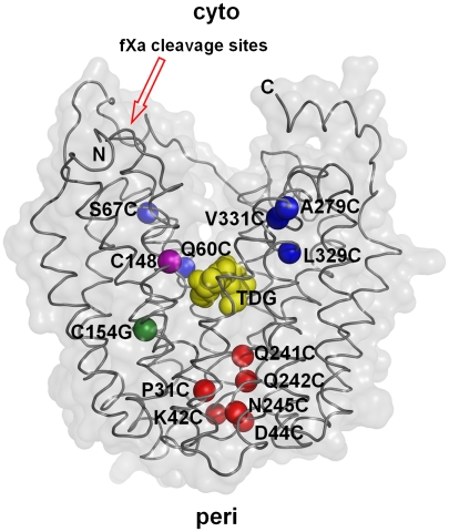
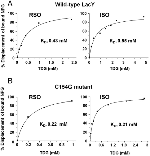
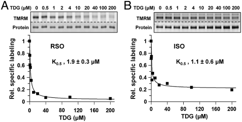
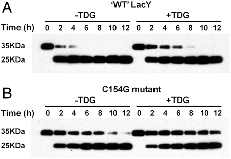
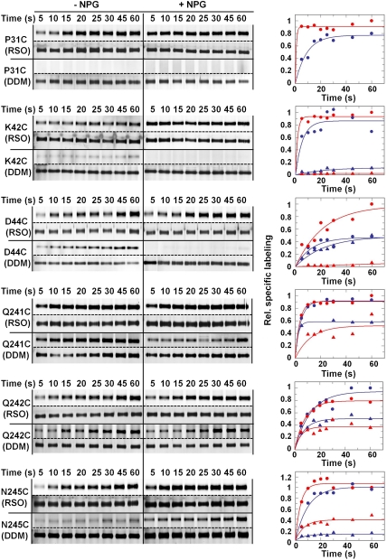
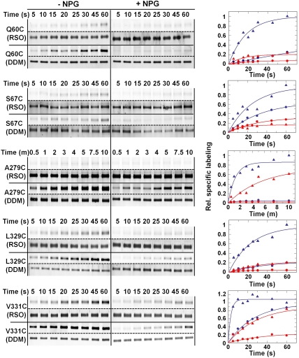
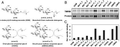
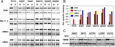
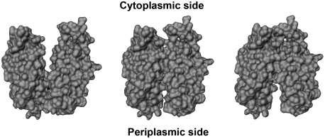
LacY mutant Cys154 → Gly exhibits a periplasmic-closed crystal structure identical to the WT, but is periplasmic-open in the membrane. The mutant hardly catalyzes transport, but binds galactosides from either side of the membrane with the same affinity and is resistant to site-directed proteolysis relative to the pseudo-WT. Site-directed alkylation was also applied to 11 single-Cys mutants in Cys154 → Gly LacY in right-side-out membrane vesicles or after solubilization and purification in dodecyl-β-D-maltopyranoside (DDM). Unlike the pseudo-WT, Cys replacements on the periplasmic side of the Cys154 → Gly mutant label rapidly in the membrane without sugar, but labeling decreases markedly after the mutant proteins are purified. Thus, Cys154 → Gly LacY likely favors a higher-energy intermediate periplasmic-open conformation in situ, but collapses to a lower-energy periplasmic-closed conformation in DDM after purification. Notably, branched-chain or neopentyl glycol maltoside detergents stabilize Cys154 → Gly LacY in the membrane-embedded form.


Conflict of interest statement

The authors declare no conflict of interest.

Figures

Fig. 1.
Fig. 1.
Single-Cys replacements in C154G LacY. Positions of single-Cys replacements are shown superimposed on the backbone of C154G LacY (PDB ID: 1PV7). LacY is viewed perpendicular to the membrane with the N-terminal helix bundle on the left and C-terminal helix bundle on the right. Blue spheres represent single-Cys replacements on the cytoplasmic side: Q60C, S67C (helix II), A279C (helix VIII), L329C, and V331C (helix X); red spheres represent single-Cys replacements on the periplasmic side: P31C (helix I), K42C, D44C (helix II), Q241C, Q242C, and N245C (helix VII). The position of Gly154 is shown in green, and the position of Cys148 is shown in purple. Yellow spheres at the apex of cavity represent TDG. The tandem factor Xa protease sites are indicated with a red arrow.
Fig. 2.
Fig. 2.
TDG binding by wild type LacY and C154G LacY. Binding of TDG to RSO or ISO membrane vesicles containing wild-type LacY (A) or C154G LacY (B) was assayed by flow dialysis as described in Materials and Methods.
Fig. 3.
Fig. 3.
Protection of C154G/Cys148 LacY against alkylation by sugar binding. RSO (A) or ISO (B) membrane vesicles were incubated with a given concentration of TDG and then labeled with 40 μM TMRM for 5 min at 0 °C. K0,5 values were obtained by quantifying the labeling intensity as described in Materials and Methods. The concentration of TDG in each sample is indicated at the top of the gels.
Fig. 4.
Fig. 4.
Proteolysis of ‘WT’ LacY and C154G mutant. ‘WT’ LacY (A) or C154G LacY (B) with tandem factor Xa sites in loop IV/V were digested with factor Xa protease in the absence or presence of 10 mM TDG for given times at 0 °C and Western-blots were performed with anti-C-terminal antibody as described in Materials and Methods.
Fig. 5.
Fig. 5.
SDA of periplasmic C154G/single-Cys mutants. Single-Cys mutants C154G/Q241C, C154G/Q242C, C154G/N245C, C154G/P31C, C154G/K42C, or C154G/D44C in RSO membrane vesicles (solid circles) or after solubilization and purification in DDM (solid triangles) were incubated with 40 μM TMRM for given times at 0 °C in the absence (blue) or presence of 1 mM NPG (red). RSO membrane vesicles containing a given mutant and labeled with TMRM were solubilized and purified in DDM by monomeric avidin chromatography and then subjected to SDS-PAGE. Alternatively, protein was solubilized and purified in DDM prior to labeling with TMRM. The gels were assayed for TMRM fluorescence (bands above the dotted lines) and silver stained for protein (bands below the dotted lines). The data were treated semiquantitatively as described in Materials and Methods by setting the highest 60 s point obtained with RSO vesicles to 1.
Fig. 6.
Fig. 6.
SDA of cytoplasmic single-Cys mutants. Single-Cys mutants C154G/Q60C, C154G/S67C, C154G/A279C, C154G/L329C, or C154G/V331C were labeled in RSO membrane vesicles (solid circles) or in DDM as purified protein (solid triangles) for indicated times at 0 °C in the absence (blue) or presence of 1 mM NPG (red) as described in Fig. 5. Relative labeling was calculated and plotted as described in Materials and Methods.
Fig. 7.
Fig. 7.
SDA of periplasmic C154G/N245C in branched-chain maltoside detergents and glucose or maltose neopentyl glycols. (A) Structures of DDM, branched-chain maltoside detergents and maltose or glucose neopentyl glycols used. (B) SDA of C154G/N245C LacY in branched-chain maltoside detergents and maltose or glucose neopentyl glycols. RSO membrane vesicles containing C154G/N245C (0.1 mg total protein) were incubated with given detergents at a final concentration 20-fold above the critical micelle concentration. LacY was purified with monomeric avidin followed by labeling with 40 μM TMRM for 1 min at 0 °C in the absence of NPG. The concentrations (%; wt/vol) of detergents used in washing and chromatography were: DDM, 0.02; Mal 9_3, 0.06; Mal 10_2, 0.06; Mal 10_3, 0.06; Mal 11_1, 0.06; Mal 11_2, 0.02; Mal 11_3, 0.02; Mal 12_1, 0.02; OGNG, 0.2; DMNG, 0.02; LMNG, 0.02. The variability observed with respect to the proteins bands with each detergent is due primarily to differences in the retention of LacY on the monovalent avidin affinity columns under the conditions used for elution. (C) Relative specific labeling of C154G/N245 LacY in selected detergents. Specific labeling in each detergent was calculated by dividing the TMRM signal by the intensity of the protein band, and the specific labeling in DDM was set as 1.
Fig. 8.
Fig. 8.
SDA of periplasmic single-Cys mutants in DDM, Mal 11_2, DMNG and LMNG. (A) Single-Cys replacements P31C, K42C, D44C, Q241C, Q242C, and N245C were labeled with TMRM in RSO membrane vesicles (R) or as purified protein (P) in the given detergent. RSO membrane vesicles were solubilized with DDM, Mal 11_2, DMNG, and LMNG at a final concentration 20-times the critical micelle concentration and the single-Cys LacY mutants were purified by monomeric avidin affinity chromatography. RSO membrane vesicles or purified proteins were labeled with 40 μM of TMRM for 1 min at 0 °C in the absence of NPG. (B) Relative specific labeling of periplasmic single-Cys mutants after purification in DDM (black), Mal 11_2 (red), DMNG (yellow) or LMNG (blue). For each detergent, relative specific labeling in RSO membrane vesicles was set at 1. (C) Effect of NPG on labeling of cytoplasmic single-Cys mutants in Mal 11_2. RSO membrane vesicles containing single-Cys replacement Q60C, S67C, A279C, L329C, or V331C were solubilized with 0.12% Mal 11_2 (wt/vol), and the proteins were purified with monomeric avidin affinity chromatography. Purified LacY in 0.02% Mal 11_2 was labeled with 40 μM of TMRM for 1 min at 0 °C in the absence or presence of 1 mM NPG as indicated.
Fig. P1.
Fig. P1.
Molecular models of LacY. (Left), inward-facing conformation with no sugar bound, (Middle), an “occluded” intermediate conformation with bound sugar and (Right), outward-facing conformation with no bound sugar correspond to three conformational populations based on distance measurements from double electron-electron resonance measurements (5). Helices II and VIII, were removed to demonstrate openings on inner (facing inward toward the cell) and outer (facing outward, toward the intermembrane space) sides.

Similar articles

Cited by

References

    1. Kaback HR, Sahin-Toth M, Weinglass AB. The kamikaze approach to membrane transport. Nat Rev Mol Cell Biol. 2001;2:610–620. - PubMed
    1. Guan L, Kaback HR. Lessons from lactose permease. Annu Rev Biophys Biomol Struct. 2006;35:67–91. - PMC - PubMed
    1. Viitanen P, Newman MJ, Foster DL, Wilson TH, Kaback HR. Purification, reconstitution, and characterization of the lac permease of Escherichia coli. Methods Enzymol. 1986;125:429–452. - PubMed
    1. Abramson J, et al. Structure and mechanism of the lactose permease of Escherichia coli. Science. 2003;301:610–615. - PubMed
    1. Mirza O, Guan L, Verner G, Iwata S, Kaback HR. Structural evidence for induced fit and a mechanism for sugar/H(+) symport in LacY. Embo J. 2006;25:1177–1183. - PMC - PubMed

Publication types

MeSH terms

LinkOut - more resources