

Composition of incubation solution impacts in vitro protein uptake to silicone hydrogel contact lenses
- PMID: 22355245
- PMCID: PMC3283214
Composition of incubation solution impacts in vitro protein uptake to silicone hydrogel contact lenses
Abstract
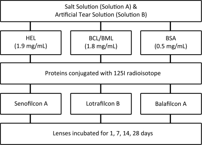
Purpose: To determine the impact of incubation solution composition on protein deposition to silicone hydrogel (SH) contact lenses using a simplistic and a complex model of the tear film.
Methods: Three SH materials--senofilcon A (SA), lotrafilcon B (LB), and balafilcon A (BA)--were incubated in two different solutions; Solution A was a simplistic augmented buffered saline solution containing a single protein, whereas Solution B was a complex artificial tear solution (ATS), containing the augmented buffered saline solution in addition to proteins, lipids, and mucins (pH=7.4). The proteins of interest (lysozyme, lactoferrin, albumin) were radiolabeled with Iodine-125 (2% protein of interest) and the accumulation of the conjugated protein to the lens materials was determined after 1, 7, 14, and 28 days of incubation. Protein deposition was measured using a gamma counter and the raw data were translated into absolute amounts (µg/lens) via extrapolation from standards.
Results: After 28 days, lysozyme uptake was significantly lower on BA lenses when incubated in Solution A (33.7 μg) compared to Solution B (56.2 μg), p<0.001. SA lenses deposited similar amounts of lysozyme when incubated in either Solution A (2.6 μg) or Solution B (4.1 μg), p>0.05. LB lenses also deposited similar amounts of lysozyme for both solutions (Solution A: 5.0 μg, Solution B: 4.7 μg, p>0.05). After 28 days, BA lenses accumulated approximately twice the amount of lactoferrin than the other lens materials, with 30.3 μg depositing when exposed to Solution A and 22.0 μg with Solution B. The difference between the two solutions was statistically significant (p<0.001). LB materials deposited significantly greater amounts of lactoferrin when incubated in Solution A (16.6 μg) compared to Solution B (10.3 μg), p<0.001. Similar amounts of lactoferrin were accumulated onto SA lenses regardless of incubation solution composition (Solution A: 8.2 μg, Solution B: 11.2 μg, p>0.05). After 28 days, albumin deposition onto BA lenses was significantly greater when lenses were incubated in Solution B (1.7 μg) compared to Solution A (0.9 μg), p<0.001. Similar amounts of albumin were deposited on SA lenses when incubated in either solution (0.6 μg versus 0.7 μg, p>0.05). LB lenses incubated in Solution A deposited more albumin compared to Solution B (0.9 μg versus 0.6 μg), p=0.003.
Discussion: Protein deposition onto SH materials varied when contact lenses were incubated in either a complex ATS compared to a single protein solution. More lysozyme accumulated onto BA lenses incubated in a complex analog of the human tear film, whereas lactoferrin deposited onto SA lenses independent of incubation solution composition. To better mimic the ex vivo environment, future studies should use more appropriate analogs of the tear film.
Figures


Similar articles
-
Impact of tear film components on lysozyme deposition to contact lenses.Optom Vis Sci. 2012 Apr;89(4):392-400. doi: 10.1097/OPX.0b013e31824c0c4a. Optom Vis Sci. 2012. PMID: 22388670
-
Impact of tear film components on the conformational state of lysozyme deposited on contact lenses.J Biomed Mater Res B Appl Biomater. 2013 Oct;101(7):1172-81. doi: 10.1002/jbm.b.32927. Epub 2013 Apr 6. J Biomed Mater Res B Appl Biomater. 2013. PMID: 23564739
-
Comparative study of lens solutions' ability to remove tear constituents.Optom Vis Sci. 2014 Sep;91(9):1045-61. doi: 10.1097/OPX.0000000000000340. Optom Vis Sci. 2014. PMID: 25105687
-
Biological and Clinical Implications of Lysozyme Deposition on Soft Contact Lenses.Optom Vis Sci. 2015 Jul;92(7):750-7. doi: 10.1097/OPX.0000000000000615. Optom Vis Sci. 2015. PMID: 26002002 Free PMC article. Review.
-
Tear analysis in contact lens wearers.Trans Am Ophthalmol Soc. 1985;83:501-45. Trans Am Ophthalmol Soc. 1985. PMID: 3914131 Free PMC article. Review.
Cited by
-
Improvement of contact lens-associated dry eye disease with the use of hydrogen peroxide.PeerJ. 2024 Dec 6;12:e18482. doi: 10.7717/peerj.18482. eCollection 2024. PeerJ. 2024. PMID: 39655327 Free PMC article.
References
-
- Jones L. Modern contact lens materials: A clinical performance update. Contact Lens Spectrum. 2002;17:24–35.
-
- Covey M, Sweeney DF, Terry R, Sankaridurg PR, Holden BA. Hypoxic effects on the anterior eye of high-Dk soft contact lens wearers are negligible. Optom Vis Sci. 2001;78:95–9. - PubMed
-
- Morgan PB, Woods CA, Tranoudis IG, Helland M, Efron N, Grupcheva CN, Jones D, Tan KO, Pesinova A, Ravn O, Santodomingo-Rubido J, Malet F, Raguz EN, Hreinsson HI, Itoi M, Chu S, Montani G, Bendoriene J, van der Worp E, Awasthi S, Lam AKC, Gonzalez-Meijome JM, Radu S, Belousov V, Gustaffson J, Stabuc SM, Hsiao JC, Nichols JJ. International Contact Lens Prescribing in 2010. Contact Lens Spectrum. 2011;26:30–5.
-
- Morgan PB, Efron N, Woods CA, Jones D, Pesinova A, Grein H-J, Tranoudi I, Chandrinos A, Itoi M, van der Worp E, Phillips G, Belousov V, Helland M, Thunholm-Hendriksson I-L, Ong A, Hung LK, Barr JT. International Contact Lens Prescribing in 2005. Contact Lens Spectrum. 2006;21:35–9.
-
- Zhao Z, Carnt NA, Aliwarga Y, Wei X, Naduvilath T, Garrett Q, Korth J, Willcox MD. Care regimen and lens material influence on silicone hydrogel contact lens deposition. Optom Vis Sci. 2009;86:251–9. - PubMed
Publication types
MeSH terms
Substances
LinkOut - more resources
Full Text Sources