. 2012:18:362-71.
Epub 2012 Feb 7.

Phenotype-genotype correlations in patients with TGFBI-linked corneal dystrophies in Taiwan

Affiliations

Phenotype-genotype correlations in patients with TGFBI-linked corneal dystrophies in Taiwan

Yu-Chih Hou et al. Mol Vis. 2012.

Abstract

Purpose: To determine the phenotype-genotype correlations in patients with corneal dystrophies associated with human transforming growth factor-β-induced (TGFBI) mutations at the National Taiwan University Hospital.

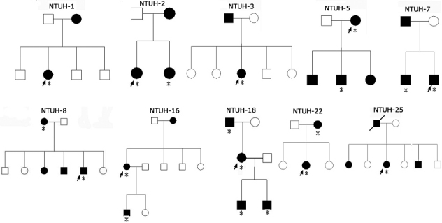
Methods: Twenty-five affected patients from 15 families with corneal dystrophies were recruited. They underwent slit-lamp biomicroscopy and visual acuity examinations. Genomic DNA was extracted from their peripheral blood, and the exons amplified from TGFBI were sequenced.

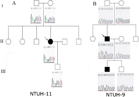
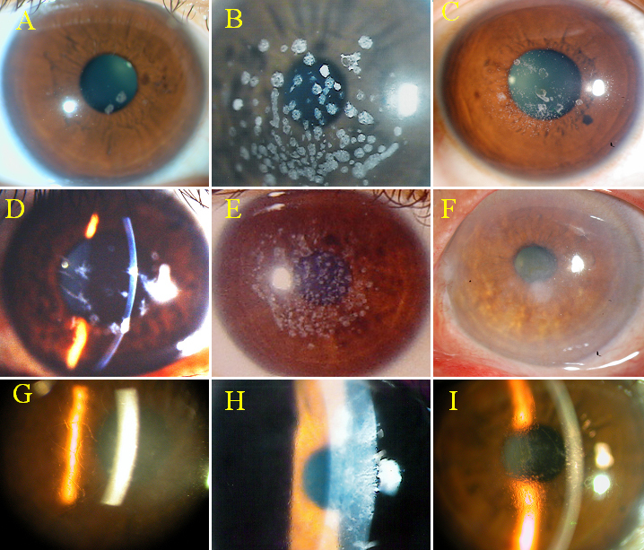
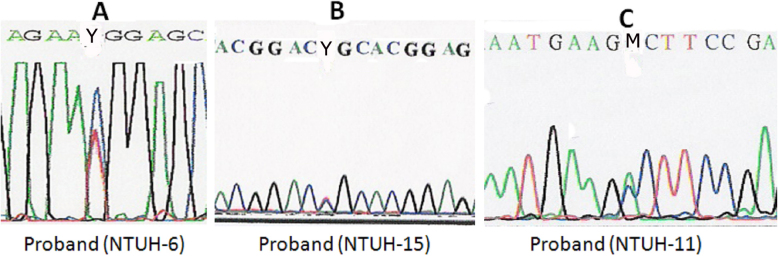
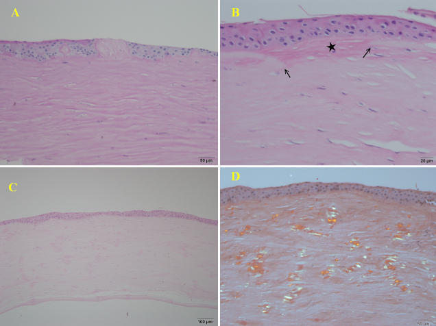
Results: Eleven patients from 9 families with granular corneal dystrophy (GCD) presented with a wide spectrum of dot or fleck opacities and shared some similar clinical features. Genetic studies revealed an R124H mutation in 5 families and an R555W mutation in 4 families. A patient with GCD type 2 and an R124H mutation showed a marked increase in opacities in the laser-assisted in situ keratomileusis (LASIK) flap interface. Six patients from 3 families with superficial honeycomb opacities had an R555Q mutation. Of the 4 patients from 3 families with variant lattice line opacities, 3 from 2 families had an R124C mutation, whereas 1 from the third family had an A546D mutation. Spontaneous mutations were detected in 2 families: an R124C mutation in 1 family with lattice corneal dystrophy (LCD) type I and an A546D mutation in the other with atypical LCD.

Conclusions: In most cases, TGFBI-linked corneal dystrophies had good phenotype-genotype correlations; however, some phenotypic variation was present. The most common mutations in Taiwan were R124H in GCD type 2 and R555W in GCD type 1. The R555Q mutation in Thiel-Behnke corneal dystrophy is not as rare in Taiwan as it is in other Asian countries. Sequencing of TGFBI can aid in the precise classification of these corneal dystrophies.


Figures

Figure 1
Figure 1
Pedigree of families with TGFBI-linked corneal dystrophies. Squares and circles represent male and female participants, respectively. Open symbols indicate unaffected individuals, and solid symbols indicate affected members. Probands are marked by an arrow. Asterisks indicate the members who underwent clinical examination and genetic analysis.
Figure 2
Figure 2
Pedigrees of the NTUH-11 and NTUH-9 families with TGFBI sequences. Open symbols indicate unaffected individuals, and solid symbols indicate affected members. Probands are marked by an arrow. A: The proband of the NTUH-11 family had a A546D mutation in exon 12, but her parents and son had normal TGFBI sequences. B: The proband of the NTUH-9 family and his elder son had a heterozygous C→T transition (R124C) in exon 4, but the proband’s parents and his younger son had TGFBI sequences without this R124 mutation.
Figure 3
Figure 3
Clinical phenotypes of TGFBI corneal dystrophies. A: Four round white opacities in the proband of the NTUH-2 family. B: Numerous crumb-shaped opacities in the proband of the NTUH-1 family. C: Numerous sand-like opacities with some rod-dot granules in the LASIK flap interface in the proband’s younger sister in the NTUH-2 family. D: Some dots with thin lines in the proband of the NTUH-6 family. E: Superficial breadcrumb-like opacities in the proband of the NTUH-4 family. F: Superficial reticular opacities in the proband of the NTUH-18 family. G: Flake-dot opacities with lattice-line opacities in the proband of the NTUH-9 family. H: Superficially central diffuse haze with some very fine and short lines in the periphery in the proband of the NTUH-15 family. I: Numerous small, polymorphic dots with some filamentous lines in the proband of the NTUH-11 family.
Figure 4
Figure 4
TGFBI mutations in 3 families with corneal dystrophies. A: A heterozygous C→T transition (R555W) in exon 12 in the proband of the NTUH-6 family. B: A heterozygous C→T transition (R124C) in exon 4 in the proband of the NTUH-15 family. C: A heterozygous C→A transition (A546D) in exon 12 in the proband of the NTUH-11 family.
Figure 5
Figure 5
Histopathology. A: The corneal specimen from the proband of the NTUH-18 family showed irregular thickness of the epithelium, vacuolization in the basal epithelium, and focal subepithelial fibrosis interposed between the irregular epithelium with a “sawtooth-like” configuration (PAS staining, 200×). B: Focal disruption of Bowman’s membrane (arrows) was replaced by subepithelial fibrotic tissue (star) (H&E staining, 400×). C: The specimen from the proband of the NTUH-11 family showed several eosinophilic deposits interspersed within the entire corneal stromal layer (H&E staining, 200×). D: These deposits showed green birefringence under a polarized microscope (Congo red staining, 200×).

Similar articles

Cited by

References

    1. Stone EM, Mathers WD, Rosenwasser GO, Holland EJ, Folberg R, Krachmer JH, Nichols BE, Gorevic PD, Taylor CM, Streb LM, Fishbaugh JA, Daley TE, Sucheski BM, Sheffield VC. Three autosomal dominant corneal dystrophies map to chromosome 5q. Nat Genet. 1994;6:47–51. - PubMed
    1. Garner A. Histochemistry of corneal granular dystrophy. Br J Ophthalmol. 1969;53:799–807. - PMC - PubMed
    1. Kivelä T, Tarkkanen A, McLean I, Ghiso J, Frangione B, Haltia M. Immunohistochemical analysis of lattice corneal dystrophies types I and II. Br J Ophthalmol. 1993;77:799–804. - PMC - PubMed
    1. Folberg R, Alfonso E, Croxatto JO, Driezen NG, Panjwani N, Laibson PR, Boruchoff SA, Baum J, Malbran ES, Fernandez-Meijide R. Clinically atypical granular corneal dystrophy with pathologic features of lattice-like amyloid deposits. A study of these families. Ophthalmology. 1988;95:46–51. - PubMed
    1. Klintworth GK. Advances in the molecular genetics of corneal dystrophies. Am J Ophthalmol. 1999;128:747–54. - PubMed

Publication types

Substances

Supplementary concepts

LinkOut - more resources