Transmission of Synucleinopathies in the Enteric Nervous System of A53T Alpha-Synuclein Transgenic Mice
- PMID: 22355263
- PMCID: PMC3268152
- DOI: 10.5607/en.2011.20.4.181
Transmission of Synucleinopathies in the Enteric Nervous System of A53T Alpha-Synuclein Transgenic Mice
Abstract
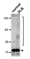
Parkinson's disease (PD) and dementia with Lewy bodies (DLB) are characterized by abnormal deposition of α-synuclein aggregates in many regions of the central and peripheral nervous systems. Accumulating evidence suggests that the α-synuclein pathology initiates in a few discrete regions and spreads to larger areas in the nervous system. Recent pathological studies of PD patients have raised the possibility that the enteric nervous system is one of the initial sites of α-synuclein aggregation and propagation. Here, we evaluated the induction and propagation of α-synuclein aggregates in the enteric nervous system of the A53T α-synuclein transgenic mice after injection of human brain tissue extracts into the gastric walls of the mice. Western analysis of the brain extracts showed that the DLB extract contained detergent-stable α-synuclein aggregates, but the normal brain extract did not. Injection of the DLB extract resulted in an increased deposition of α-synuclein in the myenteric neurons, in which α-synuclein formed punctate aggregates over time up to 4 months. In these mice, inflammatory responses were increased transiently at early time points. None of these changes were observed in the A53T mice injected with saline or the normal brain extract, nor were these found in the wild type mice injected with the DLB extract. These results demonstrate that pathological α-synuclein aggregates present in the brain of DLB patient can induce the aggregation of endogenous α-synuclein in the myenteric neurons in A53T mice, suggesting the transmission of synucleinopathy lesions in the enteric nervous system.
Keywords: Lewy body; Parkinson's disease; dementia with Lewy bodies; enteric nervous system; inflammation; protein aggregation.
Figures


References
-
- Litvan I, Bhatia KP, Burn DJ, et al. Movement Disorders Society Scientific Issues Committee report: SIC Task Force appraisal of clinical diagnostic criteria for Parkinsonian disorders. Mov Disord. 2003;18:467–486. - PubMed
-
- Dickson DW, Fujishiro H, Orr C, et al. Neuropathology of non-motor features of Parkinson disease. Parkinsonism Relat Disord. 2009;15(Suppl 3):S1–S5. - PubMed
-
- Dauer W, Przedborski S. Parkinson's disease: mechanisms and models. Neuron. 2003;39:889–909. - PubMed
-
- Polymeropoulos MH, Lavedan C, Leroy E, et al. Mutation in the alpha-synuclein gene identified in families with Parkinson's disease. Science. 1997;276:2045–2047. - PubMed
-
- Krüger R, Kuhn W, Müller T, et al. Ala30Pro mutation in the gene encoding alpha-synuclein in Parkinson's disease. Nat Genet. 1998;18:106–108. - PubMed
LinkOut - more resources
Full Text Sources
Other Literature Sources
Molecular Biology Databases

