Comparative Study
. 2012;7(2):e30885.
doi: 10.1371/journal.pone.0030885. Epub 2012 Feb 15.

iTRAQ identification of candidate serum biomarkers associated with metastatic progression of human prostate cancer

Affiliations
Comparative Study

iTRAQ identification of candidate serum biomarkers associated with metastatic progression of human prostate cancer

Ishtiaq Rehman et al. PLoS One. 2012.

Abstract

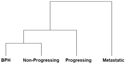
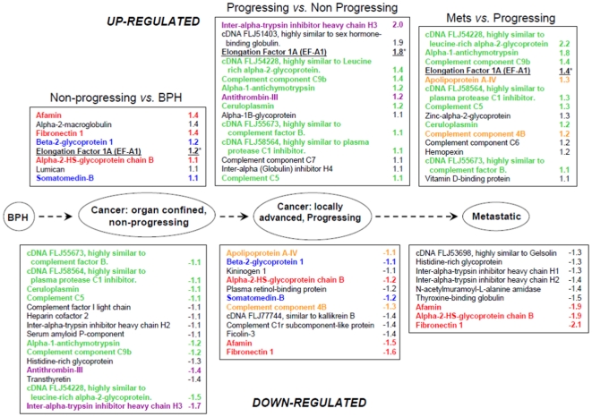
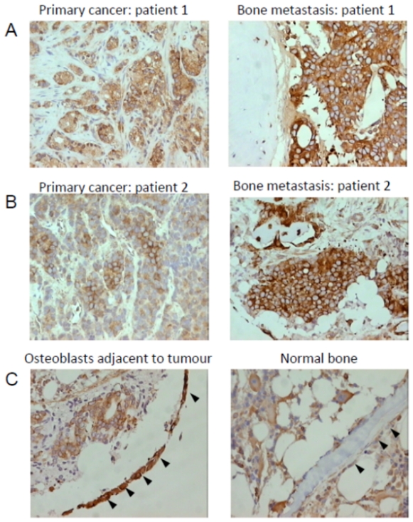
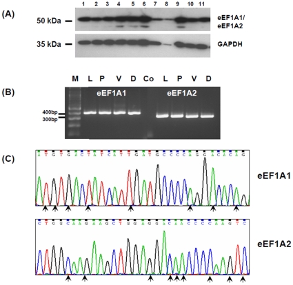
A major challenge in the management of patients with prostate cancer is identifying those individuals at risk of developing metastatic disease, as in most cases the disease will remain indolent. We analyzed pooled serum samples from 4 groups of patients (n = 5 samples/group), collected prospectively and actively monitored for a minimum of 5 yrs. Patients groups were (i) histological diagnosis of benign prostatic hyperplasia with no evidence of cancer 'BPH', (ii) localised cancer with no evidence of progression, 'non-progressing' (iii) localised cancer with evidence of biochemical progression, 'progressing', and (iv) bone metastasis at presentation 'metastatic'. Pooled samples were immuno-depleted of the 14 most highly abundant proteins and analysed using a 4-plex iTRAQ approach. Overall 122 proteins were identified and relatively quantified. Comparisons of progressing versus non-progressing groups identified the significant differential expression of 25 proteins (p<0.001). Comparisons of metastatic versus progressing groups identified the significant differential expression of 23 proteins. Mapping the differentially expressed proteins onto the prostate cancer progression pathway revealed the dysregulated expression of individual proteins, pairs of proteins and 'panels' of proteins to be associated with particular stages of disease development and progression. The median immunostaining intensity of eukaryotic translation elongation factor 1 alpha 1 (eEF1A1), one of the candidates identified, was significantly higher in osteoblasts in close proximity to metastatic tumour cells compared with osteoblasts in control bone (p = 0.0353, Mann Whitney U). Our proteomic approach has identified leads for potentially useful serum biomarkers associated with the metastatic progression of prostate cancer. The panels identified, including eEF1A1 warrant further investigation and validation.


Conflict of interest statement

Competing Interests: We are grateful to Dr. Nicholas Hoyle at Roche Diagnostics, Penzberg, Germany for his support towards consumables and reagents for the project. This does not alter the authors' adherence to all the PLoS ONE policies on sharing data and materials.

Figures

Figure 1
Figure 1. Hierarchical cluster analysis of the 4 patient groups studied.
Samples were clustered based on the similarity of their protein expression profiles observed in log10 of the iTRAQ ratios and a dendrogram generated to indicate the relationship between the samples. Squared Euclidean distance between clusters (single linkage) is shown. Varying lengths of the branch points indicate the degree of similarity; the shorter the branch the higher the degree of similarity.
Figure 2
Figure 2. Proteins showing significant differential expression (up-regulated and down-regulated) according to disease progression.
The list of differentially expressed proteins shown are based on comparisons between non-progressing versus BPH; progressing versus non-progressing and metastatic versus progressing groups. Note the differential expression of proteins either individually (black font), as pairs (blue, orange and purple fonts), or as a panel (≥3 proteins, green and red fonts). (*) = identified as a single high confidence peptide.
Figure 3
Figure 3. Representative images showing the immunoexpression of eEF1A1 in prostatic tissue, bone metastatic lesions and control bone.
(A), eEF1A1 immunoexpression can be seen in the malignant cells of the primary cancer (staining intensity = 2), and in the matched bone metastatic lesion from Patient 1 (staining intensity = 3). (B), eEF1A1 immunoexpression in the malignant cells of the primary cancer (staining intensity = 1), and in the matched bone metastatic lesion from Patient 2 (staining intensity = 3). (C), Intense eEF1A1 immunoexpression can be seen in the osteoblasts lining bone (staining intensity = 3, arrowheads), which are in close proximity to tumour cells, whereas osteoblasts lining normal bone show weak expression (staining intensity = 1, arrow heads).
Figure 4
Figure 4. The expression of eEF1A1 and eEF1A2 isoforms in human prostate cancer cell lines.
(A), Western blotting performed on prostate cancer cell lines using an antibody reactive to both the eEF1A1 and eEF1A2 isoforms. GAPDH was used as a loading control. Twenty-five micrograms of protein were loaded in each lane. Lanes 1–11: 1 = LNCaP; 2 = LNCaP-LN3; 3 = LNCaP-Pro5; 4 = LNCaP-C42; 5 = LNCaP-C4-2B; 6 = DuCaP; 7 = VCaP; 8 = Du145; 9 = PC-3; 10 = PC-3M; 11 = PC-3M-LN4. (B), Reverse-transcription PCR specific for the eEF1A1 and eEF1A2 isoforms, performed using mRNA extracted from prostate cancer cell lines: L = LNCaP; P = PC-3; V = VCaP; D = DuCaP. Control (Co), PCR was performed without mRNA. Relative equal expression of eEF1A1 and eEF1A2 mRNA can be seen in all cell lines tested (i.e. relative intensity ratio of 1.0, using Quantity One). (C), Sections of the DNA sequence chromatograms generated by sequencing the PCR products from the LNCaP cell line, confirming the specificity of the PCR primers used. Nucleotide bases unique to each isoform are marked by arrows.

Similar articles

Cited by

References

    1. Damber JE, Aus G. Prostate cancer. Lancet. 2008;371:1710–1721. - PubMed
    1. Gronberg H. Prostate cancer epidemiology. Lancet. 2003;361:859–864. - PubMed
    1. Freedland SJ, Humphreys EB, Mangold LA, Eisenberger M, Dorey FJ, et al. Risk of prostate cancer-specific mortality following biochemical recurrence after radical prostatectomy. JAMA. 2005;294:433–439. - PubMed
    1. Zhu X, van Leeuwen PJ, Bul M, Bangma CH, Roobol MJ, et al. Identifying and characterizing “escapes”- men who develop metastases or die from prostate cancer despite screening (ERSPC, section Rotterdam). Int J Cancer. 2011;129:2847–2854. - PubMed
    1. LaSpina M, Haas GP. Update on the diagnosis and management of prostate cancer. Can J Urol. 2008;15(Suppl 1):3–13; discussion 13. - PubMed

Publication types

MeSH terms