. 2012;7(2):e31274.
doi: 10.1371/journal.pone.0031274. Epub 2012 Feb 15.

TGFbeta family members are key mediators in the induction of myofibroblast phenotype of human adipose tissue progenitor cells by macrophages

Affiliations

TGFbeta family members are key mediators in the induction of myofibroblast phenotype of human adipose tissue progenitor cells by macrophages

Virginie Bourlier et al. PLoS One. 2012.

Abstract

Objective: The present study was undertaken to characterize the remodeling phenotype of human adipose tissue (AT) macrophages (ATM) and to analyze their paracrine effects on AT progenitor cells.

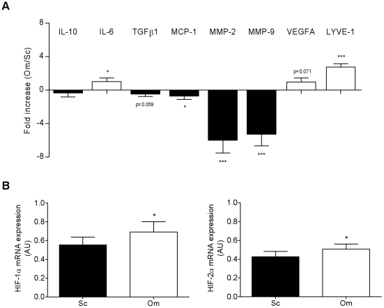
Research design and methods: The phenotype of ATM, immunoselected from subcutaneous (Sc) AT originating from subjects with wide range of body mass index and from paired biopsies of Sc and omental (Om) AT from obese subjects, was studied by gene expression analysis in the native and activated states. The paracrine effects of ScATM on the phenotype of human ScAT progenitor cells (CD34(+)CD31(-)) were investigated.

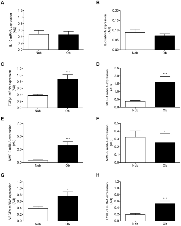
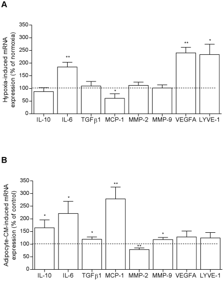
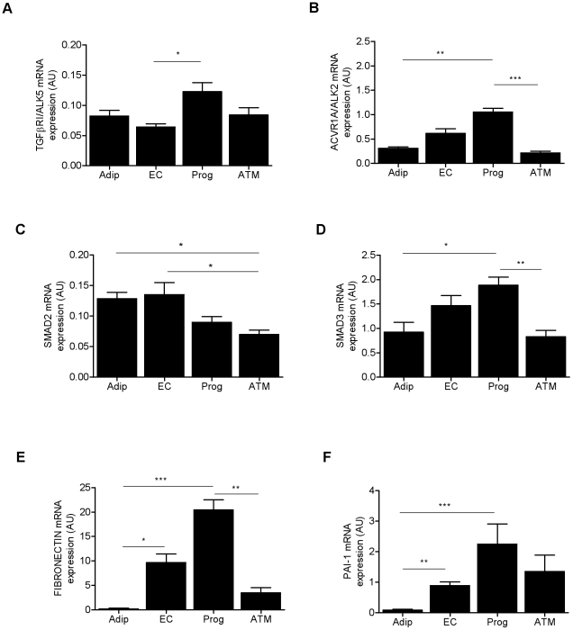
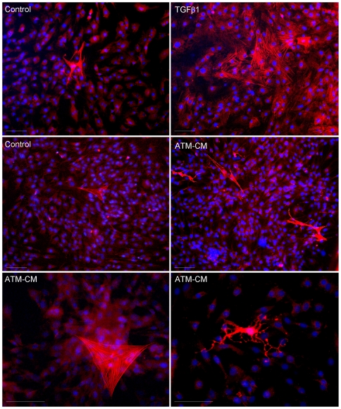
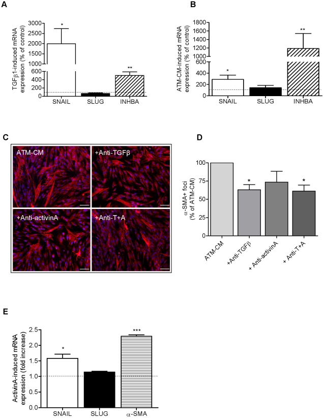
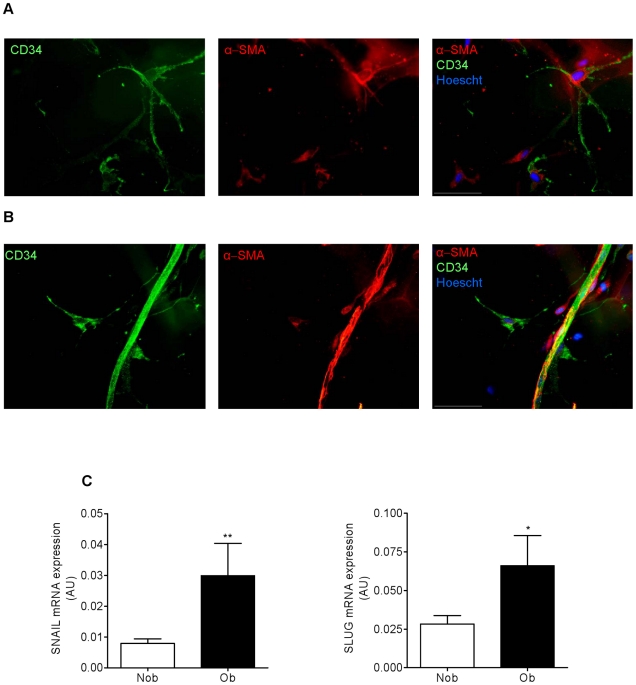
Results: Two main ATM phenotypes were distinguished based on gene expression profiles. For ScAT-derived ATM, obesity and adipocyte-derived factors favored a pro-fibrotic/remodeling phenotype whereas the OmAT location and hypoxic culture conditions favored a pro-angiogenic phenotype. Treatment of native human ScAT progenitor cells with ScATM-conditioned media induced the appearance of myofibroblast-like cells as shown by expression of both α-SMA and the transcription factor SNAIL, an effect mimicked by TGFβ1 and activinA. Immunohistochemical analyses showed the presence of double positive α-SMA and CD34 cells in the stroma of human ScAT. Moreover, the mRNA levels of SNAIL and SLUG in ScAT progenitor cells were higher in obese compared with lean subjects.

Conclusions: Human ATM exhibit distinct pro-angiogenic and matrix remodeling/fibrotic phenotypes according to the adiposity and the location of AT, that may be related to AT microenvironment including hypoxia and adipokines. Moreover, human ScAT progenitor cells have been identified as target cells for ScATM-derived TGFβ and as a potential source of fibrosis through their induction of myofibroblast-like cells.


Conflict of interest statement

Competing Interests: The authors have read the journal's policy and have the following conflicts: Research grants involving directly or indirectly (ALFEDIAM) commercial sources, Roche Diagnostic and Clarins SA. This does not alter the authors' adherence to all the PLoS ONE policies on sharing data and materials.

Figures

Figure 1
Figure 1. Obesity status affects the remodeling phenotype of human subcutaneous AT macrophages.
ATM were immunoselected from subcutaneous AT from 18 non obese individuals (Nob, mean BMI 25.4±0.6 kg/m2) and 22 obese individuals (Ob, mean BMI 43.9±1.4 kg/m2) and the transcript levels of angiogenic and matrix remodeling/fibrotic factors were determined by real-time PCR analyses. Values are means ± SEM (AU, arbitrary unit). * P<0.05, *** P<0.001 vs Nob. IL, interleukin; MCP-1, monocyte chemotactic protein 1; MMP, matrix metalloproteinase; VEGF, vascular endothelial growth factor; LYVE-1, lymphatic vessel endothelial hyaluronan receptor 1.
Figure 2
Figure 2. AT location affects the remodeling phenotype of human AT macrophages in obese individuals.
A, Transcript levels of angiogenic and matrix remodeling/fibrotic factors were determined by real-time PCR analyses of ATM immunoselected from paired biopsies of subcutaneous (Sc) and omental AT (Om). Results are expressed as fold differences between Om and Sc and are means ± SEM (n = 22 subjects, mean BMI 43.9±1.4 kg/m2). Open bars: genes up-regulated, and solid bars: genes down-regulated, in OmATM vs ScATM. B, Transcript levels of HIF-1α and -2α determined by real-time PCR analyses of ScATM and OmATM. Values are means ± SEM (AU, arbitrary unit) of the 22 paired biopsies. * P<0.05 and *** P<0.001 vs Om.
Figure 3
Figure 3. Hypoxia and mature adipocyte-derived factors affect the remodeling phenotype of human subcutaneous AT macrophages.
Transcript levels of angiogenic and matrix remodeling/fibrotic factors were determined by real-time PCR analyses on ScATM cultured for 24 h (A) in hypoxic conditions (1% O2, n = 5) and (B) in mature subcutaneous adipocyte-conditioned media (CM, n = 5). Values, expressed as a percentage of the control, are means ± SEM. * P<0.05 and ** P<0.01 vs control conditions.
Figure 4
Figure 4. Components of TGFβ-signaling pathway in human subcutaneous AT cells.
TGFβ R1 (ALK5) (A), activinA R1 (ACVR1A/ALK2) (B), SMAD2 (C), SMAD3 (D), FIBRONECTIN (E) and PAI-1 (F) transcript levels were determined by real-time PCR in ScAT mature adipocytes (Adip), endothelial cells (EC), progenitor cells (Prog) and ATM. Values are means ± SEM (AU, arbitrary units) of 5 to 27 independent. * P<0.05, ** P<0.01 and *** P<0.001 between cell types.
Figure 5
Figure 5. TGFβ1 and subcutaneous AT macrophage-conditioned media induce α-SMA expression in human AT progenitor cells.
Representative photomicrograph of immunocytochemical staining for α-SMA (red) of ScAT progenitors cells (n = 4) cultured for 48 h with basal medium (i.e, control), with TGFβ1 (5 ng/ml, n = 4, upper panel) or with ScATM-CM (n = 6, middle and lower panels). Nuclei were stained with Hoechst 33242 (blue). Magnification ×20 and ×40. White scale corresponds to 50 µm.
Figure 6
Figure 6. TGFβ1 and subcutaneous AT macrophage-conditioned media induce a myofibroblast-like phenotype in human AT progenitor cells.
A and B, Transcript levels of SNAIL, SLUG and INHBA/activinA were determined by real-time PCR analyses in ScAT progenitor cells treated for 24 h with basal medium (i.e, control, n = 4 to 5), with TGFβ1 (5 ng/ml, n = 4) or with ScATM-CM (n = 6). Results are expressed as percentage of the control and are means ± SEM. * P<0.05 and ** P<0.01 vs control media. C, Representative photomicrograph of immunocytochemical staining for α-SMA (magnification ×10) of ScAT progenitors cells (n = 3) that were cultured for 48 h with ScATM-CM (n = 5) in the presence or not of neutralizing antibodies directed against TGFβ (1 µg/ml), activinA (1 µg/ml) or both (A+T, 1 µg/ml each). White scale corresponds to 50 µm. D, Number of α-SMA+ foci per 100 nuclei. Values are expressed as a percentage of ScATM-CM and are means ± SEM. * P<0.05 vs ScATM-CM, n = 3. E, Transcript levels of SNAIL, SLUG and α-SMA were determined by real-time PCR analyses in hMADS treated or not with activinA (100 ng/ml) for 24 h (n = 4). Values are expressed as fold increase of the controland are means ± SEM. * P<0.05 and *** P<0.001 vs control media.
Figure 7
Figure 7. Obesity is associated with increased expression of myofibroblast markers in subcutaneous AT progenitor cells.
A and B, Representative photomicrograph of immunohistochemistry of whole ScAT staining: α-SMA (red), CD34 (green) and nuclei (Hoechst 33242/blue) (n = 9). White scale corresponds to 50 µm. C, SNAIL and SLUG transcript levels were determined by real-time PCR in immunoselected ScAT progenitor cells from 7 non obese (Nob) and 8 obese (Ob) individuals. Values are means ± SEM (AU, arbitrary units). * P<0.05 and ** P<0.01 vs Nob.

Similar articles

Cited by

References

    1. Weisberg SP, McCann D, Desai M, Rosenbaum M, Leibel RL, et al. Obesity is associated with macrophage accumulation in adipose tissue. J Clin Invest. 2003;112:1796–1808. - PMC - PubMed
    1. Xu H, Barnes GT, Yang Q, Tan G, Yang D, et al. Chronic inflammation in fat plays a crucial role in the development of obesity-related insulin resistance. J Clin Invest. 2003;112:1821–1830. - PMC - PubMed
    1. Curat CA, Miranville A, Sengenes C, Diehl M, Tonus C, et al. From blood monocytes to adipose tissue-resident macrophages: induction of diapedesis by human mature adipocytes. Diabetes. 2004;53:1285–1292. - PubMed
    1. Cancello R, Tordjman J, Poitou C, Guilhem G, Bouillot JL, et al. Increased infiltration of macrophages in omental adipose tissue is associated with marked hepatic lesions in morbid human obesity. Diabetes. 2006;55:1554–1561. - PubMed
    1. Lumeng CN, Bodzin JL, Saltiel AR. Obesity induces a phenotypic switch in adipose tissue macrophage polarization. J Clin Invest. 2007;117:175–184. - PMC - PubMed

Publication types

MeSH terms