. 2012;7(2):e31344.
doi: 10.1371/journal.pone.0031344. Epub 2012 Feb 15.

A MAP6-related protein is present in protozoa and is involved in flagellum motility

Affiliations

A MAP6-related protein is present in protozoa and is involved in flagellum motility

Denis Dacheux et al. PLoS One. 2012.

Abstract

In vertebrates the microtubule-associated proteins MAP6 and MAP6d1 stabilize cold-resistant microtubules. Cilia and flagella have cold-stable microtubules but MAP6 proteins have not been identified in these organelles. Here, we describe TbSAXO as the first MAP6-related protein to be identified in a protozoan, Trypanosoma brucei. Using a heterologous expression system, we show that TbSAXO is a microtubule stabilizing protein. Furthermore we identify the domains of the protein responsible for microtubule binding and stabilizing and show that they share homologies with the microtubule-stabilizing Mn domains of the MAP6 proteins. We demonstrate, in the flagellated parasite, that TbSAXO is an axonemal protein that plays a role in flagellum motility. Lastly we provide evidence that TbSAXO belongs to a group of MAP6-related proteins (SAXO proteins) present only in ciliated or flagellated organisms ranging from protozoa to mammals. We discuss the potential roles of the SAXO proteins in cilia and flagella function.


Conflict of interest statement

Competing Interests: The authors have declared that no competing interests exist.

Figures

Figure 1
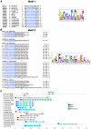
Figure 1. Identification of SAXO proteins, a MAP6-related protein family.
A. Motif 1. The N-terminal domain and its cysteine consensus sequence. Left panel: alignment of the N-terminal sequences of the proteins used for the MEME analysis in C. The boxed sequences correspond to motif 1. Amino acids corresponding to the regular expression of motif 1 are shown in blue. Right panel: motif 1 is represented as a position-specific probability matrice derived from the MEME analysis in C. B. Motif 2. Mn domains in mouse Map6-1, Map6d1, and Mn-like domains and inter-repeats in mouse Saxo1, Plasmodium SAXO and Trypanosoma SAXO. Left panel: characters in blue correspond to the regular expression of the Mn and Mn-like domains identified by the MEME analysis in C; the Mn-like domains identified manually are in italics. The underlined sequences in Map6-1 and Map6d1 correspond to the experimentally identified Mn domains in mouse , . CP motifs are boxed. IR: inter-repeat regions. Right panel: the Mn-like regular expression is represented as position-specific probability matrix derived from MEME analysis in C. C. Identification of a family of proteins containing Mn-Like domains. MEME analysis using mouse Map6s, mouse Saxo1, and only protozoan SAXO sequences identified a characteristic N-terminal motif (motif 1, dark blue boxes) in SAXO proteins and Mn-like domains (motif 2, light blue boxes) common to the SAXO and MAP6 proteins. Manually identified supplementary motifs 2 are in grey boxes (Motif 2 manual). The asterisk indicates a conserved CP sequence in the last Mn-like domain.
Figure 2
Figure 2. TbSAXO is a cytoskeleton protein in T. brucei.
A. Schematic diagram of a T. brucei cell in interphase. (N) nucleus, (K) a single kinetoplast, (F) a single flagellum, (A) a single axoneme, and PFR the single paraflagellar rod. Note that the distal end of the flagellum emerges from the flagellar pocket located at the posterior end of the cell and is then external to the cell body. B. Specificity of the polyclonal serum and the monoclonal antibody mAb25 for TbSAXO. Pre-immune and immune sera were tested on 1.106 PCF and BSF cells and 40 ng of the purified recombinant Histidine-tagged TbSAXO (His-SAXO) demonstrating the specificity of the immune serum. The derived monoclonal antibody mAb25 is also specific to TbSAXO as shown on PCF and BSF cells, histidine-tagged recombinant protein as well as PCF cells overexpressing a GFP-tagged TbSAXO (calculated MW 57.3 kD). The loading control was done with the L8C4 antibody, which identifies PFR2 protein. C. The monoclonal antibody mAb25 identifies TbSAXO on 3.106 PCF whole cells (WC), cytoskeletons (CK) and flagella (FG). The membranes were probed simultaneously with mAb25 and the loading control anti-PFR2.
Figure 3
Figure 3. TbSAXO is an axoneme-associated protein in T. brucei.
A. Immuno-labeling and localization of TbSAXO on PCF cytoskeletons. Left panel: TbSAXO localization in the flagellum was identified by the mAb25 antibody (green). Labeling extends along the entire length of the flagellum from the flagellar transition zone (*, labeled with the FTZC antibody) to the distal tip. The PFR is labeled red and begins where the flagellum exits the cell (antibody L8C4). Right panel: a merge of IF and phase contrast. N: nucleus. K: kinetoplast. F: flagellum. Bar, 5 µm. B. Images of the proximal flagellar regions of PCF cytoskeletons from cells through mitosis and cytokinesis. In each row, the left panel shows the PFR and FTZC (*) (red), the center panel TbSAXO (green), and the right panel shows merged images. The cell cycle stages are defined as 1K1N1F (1 Kinetoplast, 1 Nucleus, 1 Flagellum), 1K1N2F, 2K1N2F and 2K2N2F in rows a–d respectively. TbSAXO labeling is present immediately distal to the FTZC and is clearly distinct from PFR staining. Bar, 1 µm. C. Immuno-gold electron microscopy reveals that TbSAXO is localized in the axoneme. Mab25 immuno-gold particles can be seen mainly on the axoneme and not on the PFR of flagella of PCF WT cells. Bars, 100 nm.
Figure 4
Figure 4. TbSAXO is a microtubule-associated protein and a microtubule-stabilizing protein.
Mammalian cells (U-2 OS) expressing either MAP6-1-GFP (row a), TbSAXO-Myc (row b), or various truncated versions of TbSAXO-Myc (rows c–j) (constructs are represented on the schemes on the right panel). In each case, the transfected cells were incubated at 37°C or 4°C to test for MTs cold stability. Anti-tubulin (TAT1) and anti-Myc antibodies provided the images in left and centre columns respectively at each temperature. The right columns for each temperature set are merged images. The cells were subjected to short extraction before fixation and immuno-labeling. TbSAXO MT stabilization is seen in images b, c, e, h and j. MT stabilization is also observed in the positive control MAP6-1-GFP expressing cells (a). Nuclei were labeled with DAPI. Bar, 20 µm.
Figure 5
Figure 5. The MnL domains are required for flagellum targeting and binding in T. brucei.
Immuno-localization using WB (left panels) and IF (right panels) of C-terminal, Myc-tagged, TbSAXO truncations after over-expression in PCF cells. WB panels: 3.106 whole cells (WC), cytoskeletons (CK) and flagella (FG) were loaded on a 15% SDS-PAGE gel. The membranes were probed simultaneously with L8C4 (for PFR loading control) and anti-Myc antibodies. Lanes 1–3: non-induced cells (−Tet). Lane 4–6: cells induced for over-expression (+Tet) for 24 h, except TbSAXO-1-35-Myc (c), which was induced for 48 h. Immuno-fluorescence panels: cytoskeletons from over-expressing cells were probed with anti-Myc antibody. The kinetoplasts and nuclei were labeled with DAPI. Expression of the Myc-recombinant proteins was induced for 6 h except for TbSAXO-36-213-Myc (g), which was induced for 18 h. OF: old flagellum. NF: new flagellum. MS: mitotic spindle. Nuclei and kinetoplasts were labeled with DAPI. These results indicate that the Mn domains are necessary and sufficient for targeting TbSAXO to the flagellum. Bar, 5 µm.
Figure 6
Figure 6. TbSAXO RNAi knockdowns exhibit impaired flagellar motility.
Inducible RNAiTbSAXO in PCF (a, b, c, d) and BSF (e, f, g, h) cells. Growth curves of PCF (a) and BSF (e) RNAiTbSAXO cell lines. Corresponding WBs (PCF in b, BSF in f) of WT (parental), RNAi non-induced (-), and 24 h and 96 h induced cells probed with mAb25 and L8C4 (anti-PFR2). For PCF 5.106 cells were used and 1.25×10⁵ cells for BSF. c. Sedimentation assay of PCF RNAi. WT (closed squares). RNAi non-induced (−TET) (closed triangles) and induced (+TET) (open circles). d. Mobility graph obtained from Movie S1. The positions of individual cells are plotted at 2.5 s intervals. Open circle: starting position of each cell. Arrowhead: ending position. Number in parentheses: time in seconds of a given cell was within the field of view. Bar, 10 µm. g. Graph of cell populations with orthodox and unorthodox kinetoplast number in BSF RNAi cultures (72 h of induction). K: kinetoplast. N: nucleus. Asterisks indicate statistical significance compared with the WT population, and −TET versus +TET condition (*P<0.1; ** P<0.05; ***P<0.01). h Electron-micrograph of a thin section of an aberrant BSF RNAi induced cell (72 h). (*) indicates a flagellum. Scale bar, 2 µm. Error bars in a, c, e, and g represent the standard error from 3 independent experiments.

Similar articles

Cited by

References

    1. Amos LA, Schlieper D. Microtubules and maps. Adv Protein Chem. 2005;71:257–298. - PubMed
    1. Bosc C, Andrieux A, Job D. STOP proteins. Biochemistry. 2003;42:12125–12132. - PubMed
    1. Brinkley BR, Cartwright J., Jr Cold-labile and cold-stable microtubules in the mitotic spindle of mammalian cells. Ann N Y Acad Sci. 1975;253:428–439. - PubMed
    1. Gory-Faure S, Windscheid V, Bosc C, Peris L, Proietto D, et al. STOP-like protein 21 is a novel member of the STOP family, revealing a Golgi localization of STOP proteins. J Biol Chem. 2006;281:28387–28396. - PubMed
    1. Margolis RL, Rauch CT, Job D. Purification and assay of a 145-kDa protein (STOP145) with microtubule-stabilizing and motility behavior. Proc Natl Acad Sci U S A. 1986;83:639–643. - PMC - PubMed

Publication types

MeSH terms

Substances

LinkOut - more resources