. 2012;7(2):e31525.
doi: 10.1371/journal.pone.0031525. Epub 2012 Feb 15.

Osteoadherin accumulates in the predentin towards the mineralization front in the developing tooth

Affiliations

Osteoadherin accumulates in the predentin towards the mineralization front in the developing tooth

Hero Nikdin et al. PLoS One. 2012.

Abstract

Background: Proteoglycans (PG) are known to be involved in the organization and assembly of the extracellular matrix (ECM) prior to mineral deposition. Osteoadherin (OSAD), a keratan sulphate PG is a member of the small leucine-rich (SLRP) family of PGs and unlike other SLRPs, OSAD expression is restricted to mineralized tissues. It is proposed to have a high affinity for hydroxyapatite and has been shown to be expressed by mature osteoblasts but its exact role remains to be elucidated.

Methodology/principal findings: We investigated the protein distribution of OSAD in the developing mouse tooth using immunohistochemistry and compared its expression with other SLRPs, biglycan (BGN), decorin (DCN) and fibromodulin (FMD). OSAD was found to be specifically localized in the predentin layer of the tooth and focused at the mineralization front. These studies were confirmed at the ultrastructural level using electron microscopy (iEM), where the distribution of immunogold labeled OSAD particles were quantified and significant amounts were found in the predentin, forming a gradient towards the mineralization front. In addition, iEM results revealed OSAD to lie in close association with collagen fibers, further suggesting an important role for OSAD in the organization of the ECM. The expression profile of mineralization-related SLRP genes by rat dental pulp cells exposed to mineralization inducing factors, showed an increase in all SLRP genes. Indeed, OSAD expression was significantly increased during the mineralization process, specifically following, matrix maturation, and finally mineral deposition. Alizarin Red S staining for calcium deposition showed clear bone-like nodules, which support matrix maturation and mineralization.

Conclusions: These studies provide new evidence for the role of OSAD in the mineralization process and its specific localization in the predentin layer accumulating at the mineralization front highlighting its role in tooth development.


Conflict of interest statement

Competing Interests: The authors have declared that no competing interests exist.

Figures

Figure 1
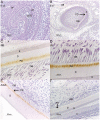
Figure 1. Immunostaining of OSAD in the developing mouse tooth.
No signal was detected for OSAD at the early bell stage of the tooth E15 (A), OSAD was first localized weakly within the alveolar bone at E17 (B), and once dentinogenesis was initiated, it continued throughout crown formation from NB (C), d5 (D) and into the adult (F). The expression of OSAD was restricted to the predentin layer in all developmental phases, Control (F) sections were incubated with OSAD antibody (0.2 µg/µl) in the presence of 10× excess of recombinant mouse OSAD protein (0.1 µg/µl). A = ameloblasts, AB = alveolar bone, D = dentin, DF = dental follicle, E = enamel, pA = pre-ameloblasts, PD = predentin, pOB = pre-odontoblasts and OB = odontoblasts.
Figure 2
Figure 2. Immunostaining of OSAD in comparison to the other SLRPs in d5 mice.
OSAD (A) expression is clearly noted in the predentin towards the predentin/dentin interface, BGN (B), DCN (C) and FMD (D) were detected again in the predentin but more closely located to the odontoblastic cell layer. A = ameloblasts, AB = alveolar bone, D = dentin, DF = dental follicle, E = enamel, pA = pre-ameloblasts, PD = predentin, pOB = pre-odontoblasts and OB = odontoblasts.
Figure 3
Figure 3. Electron images of OSAD in mouse predentin at different developmental stages.
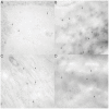
Electron images illustrating gold-labeled OSAD in the highly active predentin throughout all the different developmental stages examined (E15 (A), NB (B), D5 (C) and adult (D)). The number of gold-particles detected increased with development from E15 to NB. The elevated levels of OSAD expression were maintained in the predentin to d5 and adulthood. Localization of OSAD close to collagen fibers are very clearly observed in d5 mouse (C).
Figure 4
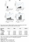
Figure 4. Quantification of OSAD expression in the developing incisor.
Quantification of the gold-labeled particles in the predentin (proximal, central and distal), dentin and enamel following ultrastructural analysis of E15, NB, d5 and adult incisors. The results are expressed as number of particles/µm2 (Au/µm2). Statistically significant differences p<0.05 are denoted by *.
Figure 5
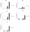
Figure 5. Gene expression of OSAD increased when induced with mineralizing media in rDPC.
rDPC were left to differentiate for 21 days in media supplemented with 50 µg/ml ascorbic acid phosphate and 10 mM β-glycerophosphate to induce a mineralizing phenotype. The changes in gene expression were measured after d3, 7, 14 and 21. Data showed an increased expression of all SLRPs (BGN, DCN, FMD and OSAD) over this period, in particular OSAD was highly expressed as the rDPC entered matrix maturation and mineralization. OCN, a mineralized tissue specific marker, also demonstrated a significant increase in expression with the onset of mineralization.
Figure 6
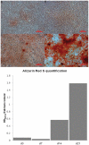
Figure 6. Alizarin Red S staining of rDPC in mineralizing culture.
Mineral deposition was examined using Alizarin Red S staining. rDPC were investigated after d3 (A), d7 (B), d14 (C) and d21 (D). Clear mineralizing nodules were observed in the osteogenically induced cultures at d21 (D). Alizarin Red S quantification demonstrated elevated calcium content by nearly 10-fold at d14 and d21 compared to d3 cultures.

Similar articles

Cited by

References

    1. Goldberg M, Septier D, Lecolle S, Chardin H, Quintana MA, et al. Dental mineralization. Int J Dev Biol. 1995;39:93–110. - PubMed
    1. Zhou HY. Proteomic analysis of hydroxyapatite interaction proteins in bone. Ann N Y Acad Sci. 2007;1116:323–326. - PubMed
    1. Embery G, Hall R, Waddington R, Septier D, Goldberg M. Proteoglycans in dentinogenesis. Crit Rev Oral Biol Med. 2001;12:331–349. - PubMed
    1. Hocking AM, Shinomura T, McQuillan DJ. Leucine-rich repeat glycoproteins of the extracellular matrix. Matrix Biol. 1998;17:1–19. - PubMed
    1. Goldberg M, Takagi M. Dentine proteoglycans: composition, ultrastructure and functions. Histochem J. 1993;25:781–806. - PubMed

Publication types

MeSH terms