

The use of fluorescence microscopy to study the association between herpesviruses and intrinsic resistance factors
- PMID: 22355446
- PMCID: PMC3280513
- DOI: 10.3390/v3122412
The use of fluorescence microscopy to study the association between herpesviruses and intrinsic resistance factors
Abstract
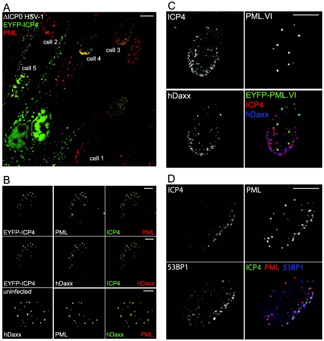
Intrinsic antiviral resistance is a branch of antiviral defence that involves constitutively expressed cellular proteins that act within individual infected cells. In recent years it has been discovered that components of cellular nuclear structures known as ND10 or PML nuclear bodies contribute to intrinsic resistance against a variety of viruses, notably of the herpesvirus family. Several ND10 components are rapidly recruited to sites that are closely associated with herpes simplex virus type 1 (HSV-1) genomes during the earliest stages of infection, and this property correlates with the efficiency of ND10 mediated restriction of HSV-1 replication. Similar but distinct recruitment of certain DNA damage response proteins also occurs during infection. These recruitment events are inhibited in a normal wild type HSV-1 infection by the viral regulatory protein ICP0. HSV‑1 mutants that do not express ICP0 are highly susceptible to repression through intrinsic resistance factors, but they replicate more efficiently in cells depleted of certain ND10 proteins or in which ND10 component recruitment is inefficient. This article presents the background to this recruitment phenomenon and summaries how it is conveniently studied by fluorescence microscopy.
Keywords: Herpes Simplex Virus type 1; ICP0; ND10; PML nuclear bodies; SUMO; intrinsic antiviral resistance.
Figures
Similar articles
-
Components of promyelocytic leukemia nuclear bodies (ND10) act cooperatively to repress herpesvirus infection.J Virol. 2013 Feb;87(4):2174-85. doi: 10.1128/JVI.02950-12. Epub 2012 Dec 5. J Virol. 2013. PMID: 23221561 Free PMC article.
-
Effect of SUMO-SIM Interaction on the ICP0-Mediated Degradation of PML Isoform II and Its Associated Proteins in Herpes Simplex Virus 1 Infection.J Virol. 2020 Jun 1;94(12):e00470-20. doi: 10.1128/JVI.00470-20. Print 2020 Jun 1. J Virol. 2020. PMID: 32295906 Free PMC article.
-
Dynamic Response of IFI16 and Promyelocytic Leukemia Nuclear Body Components to Herpes Simplex Virus 1 Infection.J Virol. 2015 Oct 14;90(1):167-79. doi: 10.1128/JVI.02249-15. Print 2016 Jan 1. J Virol. 2015. PMID: 26468536 Free PMC article.
-
Role of ND10 nuclear bodies in the chromatin repression of HSV-1.Virol J. 2016 Apr 5;13:62. doi: 10.1186/s12985-016-0516-4. Virol J. 2016. PMID: 27048561 Free PMC article. Review.
-
The Role of ND10 Nuclear Bodies in Herpesvirus Infection: A Frenemy for the Virus?Viruses. 2021 Feb 3;13(2):239. doi: 10.3390/v13020239. Viruses. 2021. PMID: 33546431 Free PMC article. Review.
Cited by
-
The Telomeric Response to Viral Infection.Viruses. 2017 Aug 9;9(8):218. doi: 10.3390/v9080218. Viruses. 2017. PMID: 28792463 Free PMC article. Review.
-
The replication defect of ICP0-null mutant herpes simplex virus 1 can be largely complemented by the combined activities of human cytomegalovirus proteins IE1 and pp71.J Virol. 2013 Jan;87(2):978-90. doi: 10.1128/JVI.01103-12. Epub 2012 Nov 7. J Virol. 2013. PMID: 23135716 Free PMC article.
-
Epigenetic control of cytomegalovirus latency and reactivation.Viruses. 2013 May 23;5(5):1325-45. doi: 10.3390/v5051325. Viruses. 2013. PMID: 23698401 Free PMC article. Review.
-
The histone chaperone HIRA promotes the induction of host innate immune defences in response to HSV-1 infection.PLoS Pathog. 2019 Mar 22;15(3):e1007667. doi: 10.1371/journal.ppat.1007667. eCollection 2019 Mar. PLoS Pathog. 2019. PMID: 30901352 Free PMC article.
-
Distinct temporal roles for the promyelocytic leukaemia (PML) protein in the sequential regulation of intracellular host immunity to HSV-1 infection.PLoS Pathog. 2018 Jan 8;14(1):e1006769. doi: 10.1371/journal.ppat.1006769. eCollection 2018 Jan. PLoS Pathog. 2018. PMID: 29309427 Free PMC article.
References
-
- Bieniasz P.D. Intrinsic immunity: A front-line defense against viral attack. Nat. Immunol. 2004;5:1109–1115. - PubMed
-
- Tavalai N., Stamminger T. New insights into the role of the subnuclear structure ND10 for viral infection. Biochim. Biophys. Acta. 2008;1783:2207–2221. - PubMed
-
- Maul G.G., Guldner H.H., Spivack J.G. Modification of discrete nuclear domains induced by herpes simplex virus type 1 immediate early gene 1 product (ICP0). J. Gen. Virol. 1993;74:2679–2690. - PubMed
Publication types
MeSH terms
Substances
Grants and funding
LinkOut - more resources
Full Text Sources
Miscellaneous
