Review
. 2012 Jan;4(1):83-101.
doi: 10.3390/v4010083. Epub 2012 Jan 13.

The curious case of arenavirus entry, and its inhibition

Affiliations
Review

The curious case of arenavirus entry, and its inhibition

Jack H Nunberg et al. Viruses. 2012 Jan.

Abstract

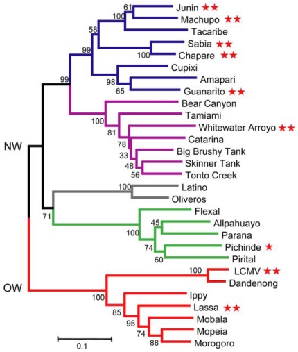
Arenaviruses comprise a diverse family of enveloped negative-strand RNA viruses that are endemic to specific rodent hosts worldwide. Several arenaviruses cause severe hemorrhagic fevers in humans, including Junín and Machupo viruses in South America and Lassa fever virus in western Africa. Arenavirus entry into the host cell is mediated by the envelope glycoprotein complex, GPC. The virion is endocytosed on binding to a cell-surface receptor, and membrane fusion is initiated in response to physiological acidification of the endosome. As with other class I virus fusion proteins, GPC-mediated membrane fusion is promoted through a regulated sequence of conformational changes leading to formation of the classical postfusion trimer-of-hairpins structure. GPC is, however, unique among the class I fusion proteins in that the mature complex retains a stable signal peptide (SSP) as a third subunit, in addition to the canonical receptor-binding and fusion proteins. We will review the curious properties of the tripartite GPC complex and describe evidence that SSP interacts with the fusion subunit to modulate pH-induced activation of membrane fusion. This unusual solution to maintaining the metastable prefusion state of GPC on the virion and activating the class I fusion cascade at acidic pH provides novel targets for antiviral intervention.

Keywords: antiviral; arenavirus; endosome; envelope glycoprotein; fusion inhibitor; fusion protein; hemorrhagic fever; pH-dependent membrane fusion; stable signal peptide; zinc binding.


Figures

Figure 1
Figure 1
Phylogenetic relationships between arenaviruses. The Old World (OW) virus lineage is depicted in red, and New World (NW) lineages are subdivided into clades: A (green), B (blue), C (gray) and a recombinant A-B lineage (purple). This phylogenetic tree is based on amino-acid comparisons of the GPC. Red stars that follow virus names indicate the ability to infect humans: ** denote lethal hemorrhagic fever viruses and lymphocytic choriomeningitis virus (LCMV), and * identifies infections with few or no clinical manifestations [1]. This image is adapted from [1] and used by permission.
Figure 2
Figure 2
Schematic representation of GPC open-reading frame, stable signal peptide (SSP) sequence alignment and the tripartite GPC protein complex. (A) The GPC open‑reading frame is diagrammed. Cleavage sites for signal peptidase (SPase) and subtilisin-like kexin protease-1/site-1-protease (SKI-1/S1P) are indicated, as are the mature SSP, G1 and G2 subunits. In SSP, the myristoylation at glycine 2 is marked, and the shaded regions denote the two hydrophobic regions (hɸ1 and hɸ2, highlighted in red in the sequence comparisons below). The transmembrane and cytoplasmic domains of G2 are indicated, as well as the two heptad‑repeat regions (shaded) and disulfide-bonded hinge region. The sequence comparison of SSP among arenaviruses is adapted from [32], in which accession numbers are listed. In addition to hɸ1 and hɸ2 (red), the conserved myristoylation motif, K33 and C57 residues are highlighted in gray. (B) Schematic drawing illustrating the subunit organization of the tripartite GPC complex. The membrane is shown in gray. SSP spans the membrane twice, and salient features are indicated: Membrane association of the myristoylated N-terminus, K33 in the SSP ectodomain, and the intersubunit zinc-binding motif that bridges the C‑terminal cytoplasmic domains of SSP and G2. The two heptad-repeat regions in the G2 ectodomain are depicted in black. The drawing is not to scale and the structural relationships among subunits is not known.
Figure 3
Figure 3
NMR structure of the zinc-binding domain in the cytoplasmic tail of G2. (A) Ribbon model of the deduced structure. Coloring changes from blue (N terminus) to red (C terminus). The zinc ions are depicted as spheres, and the coordinating cysteine and histidine sidechains are displayed. Black diamonds on the C-terminal half of the ribbon depict the two pairs of basic residues (KK and RR) implicated in ER retention/retrieval. (B) Superimposed backbone traces are colored to indicate the degree of sequence conservation among arenaviruses: highly conserved residues are shown in purple and highly variable residues in cyan.
Figure 4
Figure 4
K33 in SSP modulates pH-induced activation of membrane fusion. (A) Mutations that reduce positive polarity at SSP K33 were introduced into SSP and the membrane-fusion activity of the GPC complex was determined in our standard assay for cell-cell fusion. All SSPs were expressed in trans with the G1G2 precursor to reconstitute GPC, and fusion activity was normalized to that of the wild-type (K33) SSP. GPC and G1G2 indicate, respectively, the complete GPC open-reading frame and the precursor alone, in the absence of SSP. (B) The pH-dependence of the mutant GPC complexes was determined by varying the pH of the triggering pulse of acidic medium. The nominal optimal pH for each mutant is listed at right. (C) Model for pH-induced activation of GPC, and its inhibition by mutations at K33 and by small-molecule compounds. Acidic pH destabilizes the metastable prefusion GPC complex to favor an activated state, that spontaneously undergoes the class I structural reorganization resulting in formation of the six-helix bundle and membrane fusion. By contrast, K33 mutations and inhibitor binding disfavor the transition to the activated state. The molecular basis for maintaining the equilibrium between prefusion and activated states is unknown.
Figure 5
Figure 5
Chemically distinct classes of arenavirus-specific fusion inhibitors. Compounds from SIGA Technologies are indicated with the ST prefix [15,16,85,86] and 8C1 and 17C8 were described at The Scripps Research Institute [17]. All lead compounds were identified through high-throughput screening for inhibition of arenavirus infection or GPC‑mediated entry. The compounds all bind to a common site on prefusion GPC and act through the pH‑sensitive SSP-G2 interface, but differ in their specificities for NW and/or OW arenaviruses (as indicated).

References

    1. Charrel R.N., de Lamballerie X. Zoonotic aspects of arenavirus infections. Vet. Microbiol. 2010;140:213–220. - PubMed
    1. Palacios G., Savji N., Hui J., Travassos da Rosa A., Popov V., Briese T., Tesh R., Lipkin W.I. Genomic and phylogenetic characterization of Merino Walk virus, a novel arenavirus isolated in South Africa. J. Gen. Virol. 2010;91:1315–1324. - PMC - PubMed
    1. Coulibaly-N'Golo D., Allali B., Kouassi S.K., Fichet-Calvet E., Becker-Ziaja B., Rieger T., Olschläger S., Dosso H., Denys C., Ter Meulen J., et al. Novel arenavirus sequences in Hylomyscus sp. and Mus (Nannomys) setulosus from Côte d'Ivoire: Implications for evolution of arenaviruses in Africa. PLoS One. 2011;6:e20893. - PMC - PubMed
    1. Clegg J.C.S. Molecular phylogeny of the arenaviruses. Curr. Top. Microbiol. Immunol. 2002;262:1–24. - PubMed
    1. Salazar-Bravo J., Ruedas L.A., Yates T.L. Mammalian reservoirs of arenaviruses. Curr. Top. Microbiol. Immunol. 2002;262:25–63. - PubMed

Publication types

MeSH terms