. 2012 Jan;4(1):117-39.
doi: 10.3390/v4010117. Epub 2012 Jan 18.

Molecular and cellular aspects of rhabdovirus entry

Affiliations

Molecular and cellular aspects of rhabdovirus entry

Aurélie A V Albertini et al. Viruses. 2012 Jan.

Abstract

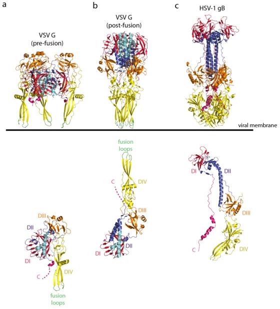
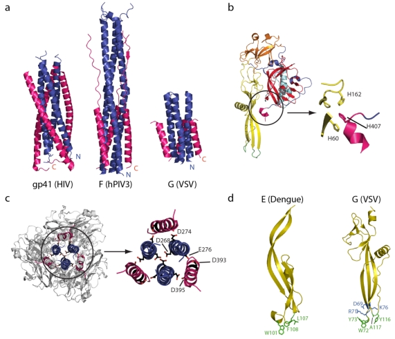
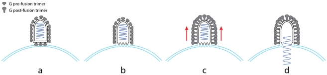
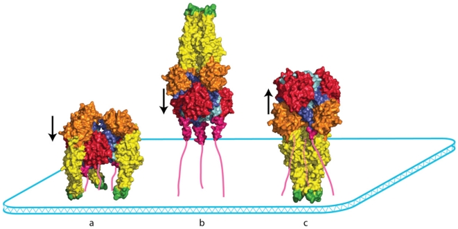
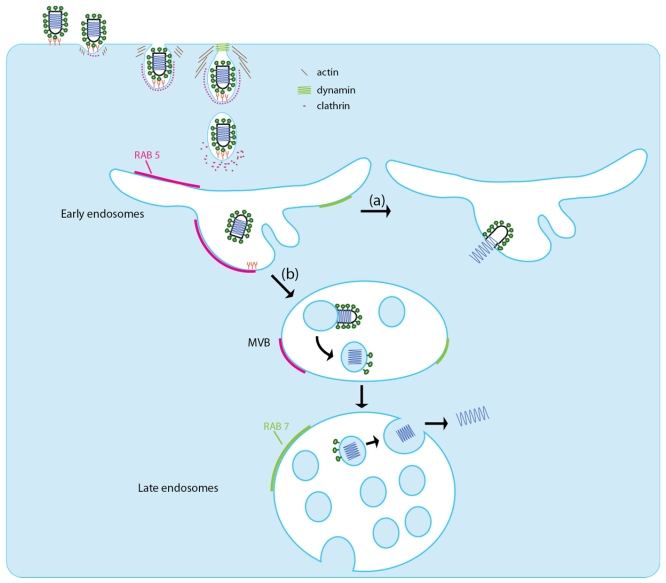
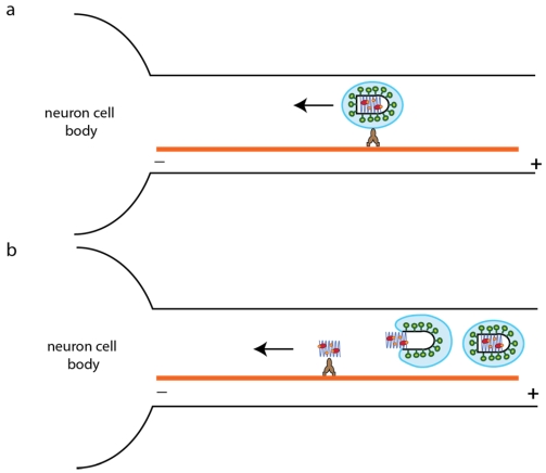
Rhabdoviruses enter the cell via the endocytic pathway and subsequently fuse with a cellular membrane within the acidic environment of the endosome. Both receptor recognition and membrane fusion are mediated by a single transmembrane viral glycoprotein (G). Fusion is triggered via a low-pH induced structural rearrangement. G is an atypical fusion protein as there is a pH-dependent equilibrium between its pre- and post-fusion conformations. The elucidation of the atomic structures of these two conformations for the vesicular stomatitis virus (VSV) G has revealed that it is different from the previously characterized class I and class II fusion proteins. In this review, the pre- and post-fusion VSV G structures are presented in detail demonstrating that G combines the features of the class I and class II fusion proteins. In addition to these similarities, these G structures also reveal some particularities that expand our understanding of the working of fusion machineries. Combined with data from recent studies that revealed the cellular aspects of the initial stages of rhabdovirus infection, all these data give an integrated view of the entry pathway of rhabdoviruses into their host cell.

Keywords: endocytosis; glycoprotein; membrane fusion; rabies virus; rhabdovirus; vesicular stomatitis virus.


Figures

Figure 1
Figure 1
(a) Ribbon diagrams of the vesicular stomatitis virus (VSV) glycoprotein (G) pre-fusion trimer (top) and protomer (bottom) (PDB code = 2J6J [73]). (b) Ribbon diagram of the VSV G post-fusion trimer (top) and protomer (bottom) (PDB code = 2CMZ [72]). G is depicted by domains; the lateral domain (domain I) is in red, the pleckstrin homology domain (domain III) is in orange, and the fusion domain (domain IV) is in yellow. The trimerization domain is colored with two shades of blue (the part of G that retains its structure during refolding is in cyan, and the part of G that is refolded during the structural transition is in deep blue). The fusion loops are green and the C-terminus is pink. (c) Ribbon diagram of the HSV-1 gB trimer (top) and protomer (bottom) (PUB: 2GUM [74]). gB is colored by domains with the same color code as VSV G. The orientation of the VSV G and HSV-1 gB trimers toward the viral membrane is indicated. The VSV G protomers are aligned on the rigid block made of the lateral domain (in red) and the cyan part of the trimerization domain. The orientation of the post-fusion protomer is thus different in the top and the bottom of the figure.
Figure 2
Figure 2
(a) A comparison of the central six-helix bundle of the class I viral fusion proteins (HIV gp41 and hPIV3 F) and class III viral fusion proteins (VSV G) in their post‑fusion conformation. The amino-terminal trimeric coiled-coil is in blue and the antiparallel lateral helices are in pink. (b) A close up view of the histidine cluster located at the fusion domain/C-terminal domain interface that has to be disrupted during the structural transition from pre- to post fusion structure. (c) Top view and close up of the VSV G trimerization domain showing the clusters of acidic residues in the post-fusion state. (d) The structural organization of the Dengue virus E fusion domain (left) and the VSV G fusion domain (right). The hydrophobic residues in the tips of the fusion domains are depicted in green and, for VSV G, charged residues that impede deep penetration of the fusion domain inside the membrane are in blue.
Figure 3
Figure 3
A model for VSV fusion [88]. In the first step (a), the flat base of VSV interacts with the target membrane most likely via activated glycoproteins that expose their fusion domain at their top. The local organization of G in this region may facilitate the formation of stalks and initial fusion pores (b). Then, a helical network of G in their post-fusion conformations is formed on the lateral part of the viral particle (c). The formation of this regular array likely induces tension in the membrane (red arrows) that drives pore enlargement and leads to complete fusion (d).
Figure 4
Figure 4
The topological problem associated with VSV G conformational changes. The VSV G pre- and post-fusion conformations are represented by a space-filling model viewed from the side. The C-terminal segments that reach the membrane are depicted as pink lines. In the pre-fusion conformation (a) and in a putative trimeric intermediate (b), the three fusion domains are located outside a pyramidal volume where the base would be the membrane and the sides would be defined by the ectodomain C-terminal segments. The converse occurs in the post-fusion state (c). Going from the pre-fusion trimer to the post‑fusion trimer is very complicated, if not impossible, without monomerization. The arrows indicate the orientation of the central helices in each conformation.
Figure 5
Figure 5
The two proposed pathways for VSV entry into the host cell. VSV binds to the host cell via an interaction with one or several unknown receptors. The VSV particles are then endocytosed in a clathrin-based, dynamin-2-dependent manner. The endocytosis vesicle is partially coated with clathrin and requires actin polymerization for efficient viral entry. Pathway (a): fusion occurs rapidly in early endosome and leads to the release of the nucleocapsid into the cytoplasm. Pathway (b): VSV fuses first with an internal vesicle inside multivesicular bodies. The nucleocapsid is then released in an internal vesicle. In the late endosome, these vesicles deliver the nucleocapsid to the cytoplasm through a back‑fusion mechanism. Small GTPase RAB5 (pink) is mainly present on early endosomes, whereas RAB7 (light green) is present mostly in late endosomes.
Figure 6
Figure 6
Two plausible pathways for rabies virus (RABV) retrograde transport in the axon. RABV may be transported by two different mechanisms on microtubules either in a vesicle (a) or as a free nucleocapsid (after membrane fusion) (b). As mentioned in the text, experimental data are in favor of pathway (a).

Similar articles

Cited by

References

    1. Rose J.K., Whitt M.A. Fields Virology. 4th. Lippincott-Raven Publishers; Philadelphia, PA, USA: 2001. Rhabdoviridae: The viruses and their replication; pp. 1221–1224.
    1. Fort P., Albertini A., Van-Hua A., Berthomieu A., Roche S., Delsuc F., Pasteur N., Capy P., Gaudin Y., Weill M. Fossil rhabdoviral sequences integrated into arthropod genomes: Ontogeny, evolution, and potential functionality. Mol. Biol. Evol. 2011 - PubMed
    1. Mebatsion T., Weiland F., Conzelmann K.K. Matrix protein of rabies virus is responsible for the assembly and budding of bullet-shaped particles and interacts with the transmembrane spike glycoprotein G. J. Virol. 1999;73:242–250. - PMC - PubMed
    1. Doms R.W., Keller D.S., Helenius A., Balch W.E. Role for adenosine triphosphate in regulating the assembly and transport of vesicular stomatitis virus G protein trimers. J. Cell Biol. 1987;105:1957–1969. - PMC - PubMed
    1. Gaudin Y., Ruigrok R.W., Tuffereau C., Knossow M., Flamand A. Rabies virus glycoprotein is a trimer. Virology. 1992;187:627–632. - PMC - PubMed

Publication types

MeSH terms