

Small intestine perforation due to metastatic uterine cervix interdigitating dendritic cell sarcoma: a rare manifestation of a rare disease
- PMID: 22355501
- PMCID: PMC3282451
- DOI: 10.4081/rt.2011.e46
Small intestine perforation due to metastatic uterine cervix interdigitating dendritic cell sarcoma: a rare manifestation of a rare disease
Abstract
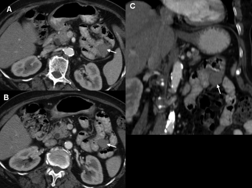
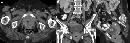
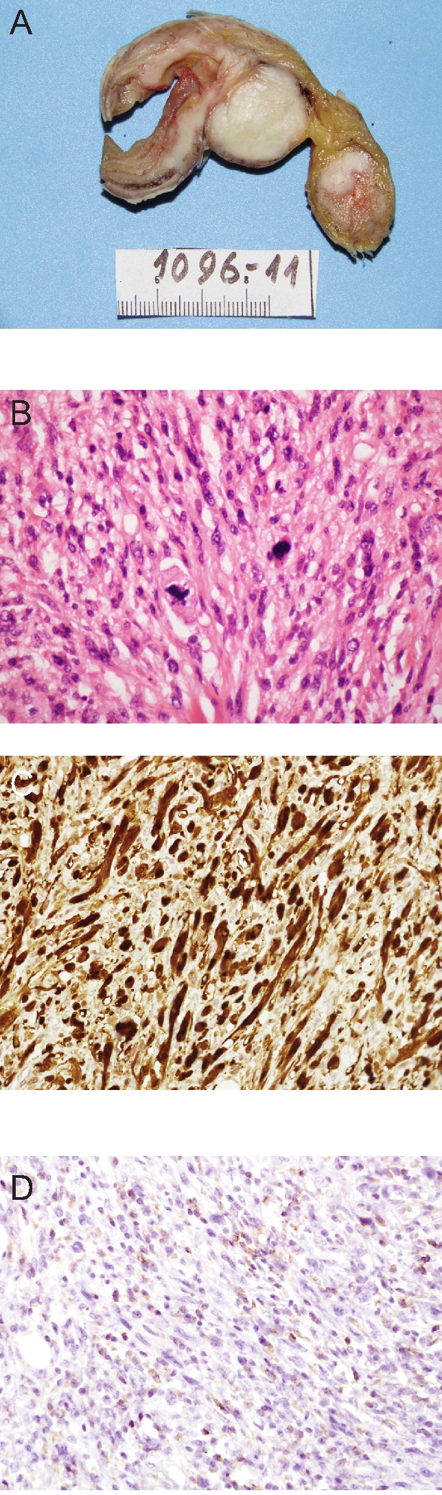
Interdigitating Dendritic Cell Sarcoma (IDCS) is an infrequent dendritic cell tumor which mainly affects the lymphatic system. Intestinal metastasis from uterine IDCS is extremely rare. Here we report a case of a 76-year-old female presenting with vaginal bleeding and acute abdomen. The final diagnosis revealed a small bowel perforation due to metastatic involvement from uterine cervix IDCS. In this paper, we report the clinical manifestation, computed tomography and histopathological findings helpful for the accurate diagnosis of this rare tumor.
Keywords: dendritic cell neoplasm; interdigitatin dendritic cell sarcoma; small intestine; uterine cervix..
Conflict of interest statement
Conflict of interest: the authors report no conflicts of interest.
Figures
Similar articles
-
Interdigitating Dendritic Cell Sarcoma of the Small Intestine Presenting as Spontaneous Hemoperitoneum - A Rare Case Report.Niger J Surg. 2021 Jan-Jun;27(1):71-74. doi: 10.4103/njs.NJS_55_19. Epub 2021 Mar 9. Niger J Surg. 2021. PMID: 34012247 Free PMC article.
-
Interdigitating dendritic cell sarcoma presenting simultaneously with acute myelomonocytic leukemia: report of a rare case and literature review.Int J Hematol. 2013 May;97(5):657-66. doi: 10.1007/s12185-013-1336-6. Epub 2013 Apr 19. Int J Hematol. 2013. PMID: 23605368 Review.
-
A Case Presentation of a Rare Pelvic Interdigitating Dendritic Cell Sarcoma.Cureus. 2024 Feb 14;16(2):e54220. doi: 10.7759/cureus.54220. eCollection 2024 Feb. Cureus. 2024. PMID: 38371440 Free PMC article.
-
An unusual case of extra-nodal interdigitating dendritic cell neoplasm: sarcoma or not?Acta Chir Belg. 2023 Dec;123(6):687-690. doi: 10.1080/00015458.2022.2102358. Epub 2022 Jul 20. Acta Chir Belg. 2023. PMID: 35833666
-
Interdigitating dendritic cell sarcoma and follicular dendritic cell sarcoma: histopathological findings for differential diagnosis.J Clin Exp Hematop. 2013;53(3):179-84. doi: 10.3960/jslrt.53.179. J Clin Exp Hematop. 2013. PMID: 24369219 Review.
Cited by
-
Interdigitating Dendritic Cell Sarcoma of the Small Intestine Presenting as Spontaneous Hemoperitoneum - A Rare Case Report.Niger J Surg. 2021 Jan-Jun;27(1):71-74. doi: 10.4103/njs.NJS_55_19. Epub 2021 Mar 9. Niger J Surg. 2021. PMID: 34012247 Free PMC article.
-
Interdigitating dendritic cell sarcoma presenting simultaneously with acute myelomonocytic leukemia: report of a rare case and literature review.Int J Hematol. 2013 May;97(5):657-66. doi: 10.1007/s12185-013-1336-6. Epub 2013 Apr 19. Int J Hematol. 2013. PMID: 23605368 Review.
-
Diffuse lesion and necrosis tied to poorer prognosis of interdigitating dendritic cell sarcoma: cases report and a pooled analysis.Sci Rep. 2017 Apr 6;7(1):667. doi: 10.1038/s41598-017-00719-2. Sci Rep. 2017. PMID: 28386111 Free PMC article. Review.
References
-
- Kairouz S, Hashash J, Kabbara W, et al. Dendritic cell neoplasms: An overview. Am J Hematol. 2007;82:924–8. - PubMed
-
- De Pas T, Spitaleri G, Pruneri G, et al. Dendritic cell sarcoma: an analytic overview of the literature and presentation of original five cases. Crit Rev Oncol Hematol. 2008;65:1–7. - PubMed
-
- Efune G, Sumer BD, Sarode VR, et al. Interdigitating dendritic cell sarcoma of the parotid gland: case report and literature review. Am J Otolaryngol. 2009;30:264–8. - PubMed
-
- Gaertner EM, Tsokos M, Derringer GA, et al. Interdigitating dendritic cell sarcoma. A report of four cases and review of the literature. Am J Clin Pathol. 2001;115:589–97. - PubMed
Publication types
LinkOut - more resources
Full Text Sources
