. 2011:1:2.
doi: 10.1038/srep00002. Epub 2011 Jun 14.

Zfp488 promotes oligodendrocyte differentiation of neural progenitor cells in adult mice after demyelination

Affiliations

Zfp488 promotes oligodendrocyte differentiation of neural progenitor cells in adult mice after demyelination

Mangala M Soundarapandian et al. Sci Rep. 2011.

Abstract

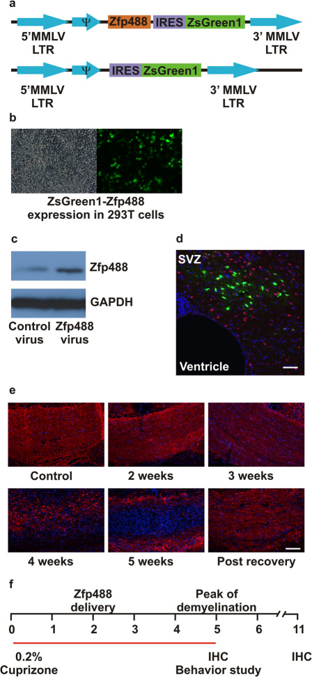
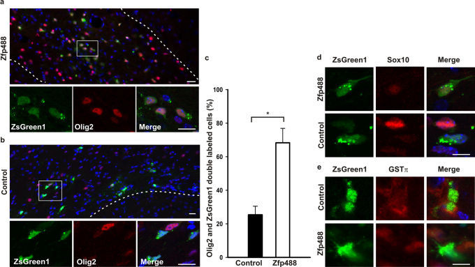
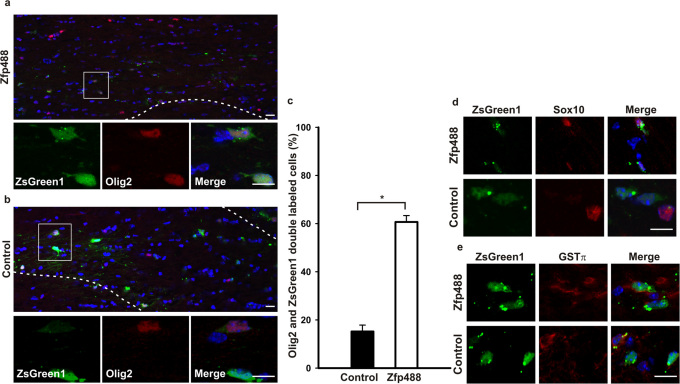
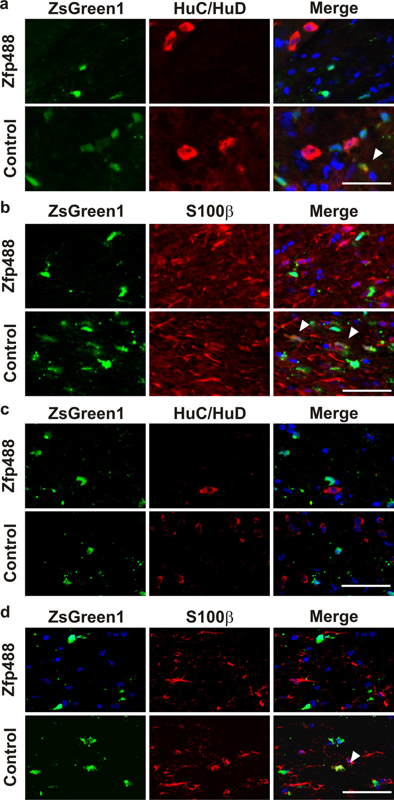
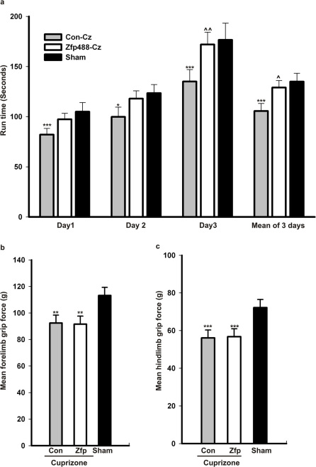
Basic helix-loop-helix transcription factors Olig1 and Olig2 critically regulate oligodendrocyte development. Initially identified as a downstream effector of Olig1, an oligodendrocyte-specific zinc finger transcription repressor, Zfp488, cooperates with Olig2 function. Although Zfp488 is required for oligodendrocyte precursor formation and differentiation during embryonic development, its role in oligodendrogenesis of adult neural progenitor cells is not known. In this study, we tested whether Zfp488 could promote an oligodendrogenic fate in adult subventricular zone (SVZ) neural stem/progenitor cells (NSPCs). Using a cuprizone-induced demyelination model in mice, we examined the effect of retrovirus-mediated Zfp488 overexpression in SVZ NSPCs. Our results showed that Zfp488 efficiently promoted the differentiation of the SVZ NSPCs into mature oligodendrocytes in vivo. After cuprizone-induced demyelination injury, Zfp488-transduced mice also showed significant restoration of motor function to levels comparable to control mice. Together, these findings identify a previously unreported role for Zfp488 in adult oligodendrogenesis and functional remyelination after injury.


Figures

Figure 1
Figure 1. Experimental design and retroviral vector construction.
(a) Schematic showing retroviral constructs, both empty vector (control) and with an insert coding for mouse Zfp488. After transduction, a Moloney murine leukemia virus (MMLV) long terminal repeats (LTR) drives Zfp488 expression. The vectors also express the fluorescent protein ZsGreen1 after an IRES to enable their identification and tracking. This promoter is not active in progenitor cells due to expression of a specific repressor element, but is transcribed once these cells start to differentiate. (b) ZsGreen1 fluorescence in Zfp488-overexpressing HEK 293T cells. (c) Western blots of Zfp488 retrovirus transduced HEK 293T lysate showing increased expression of Zfp488. (d) Representative Olig2 (red) immunolabeled image of the white matter adjoining the SVZ, 3 weeks after retrovirus injection, showing the distribution and integration of ZsGreen positive cells in mice that were not treated with cuprizone. (e) Images showing demyelination of the corpus callosum at different time points after cuprizone treatment. Sections were stained for myelin basic protein (MBP, red) and DAPI (blue). (f) For all experiments, two groups of mice were fed a 0.2% cuprizone diet for a period of 5 weeks. At two weeks, intracerebral injections into the SVZ of either control or Zfp488 expressing-retroviruses were carried out using a stereotaxic frame. After 5 weeks, mice in group I were used for behavior analysis and immunohistochemical characterization. Mice in group II were allowed to recover for an additional 6 weeks on normal diet without cuprizone. After the 6-week recovery period, tissues were collected to examine for the long-term survival of the retrovirus transduced-differentiated cells. Scale bar, 100 μm.
Figure 2
Figure 2. Zfp488 directs the differentiation of SVZ progenitor cells into mature oligodendrocytes at the peak of cuprizone-induced demyelination.
Mice on a 0.2% cuprizone diet for 2 weeks were injected with either control or Zfp488 expressing retroviruses into the SVZ. After 5 weeks in the cuprizone diet, brain sections were evaluated for the differentiation status of the virus transduced SVZ stem/progenitor cell population. (a) Representative image of the Zfp488 virus transduced SVZ-derived cells in the white matter of the corpus callosum. Higher magnification panel show that cells transduced with the Zfp488 retrovirus differentiated into oligodendrocytes and were positive for Olig2. (b) Representative image of the control (ZsGreen1) virus transduced SVZ-derived cells in the white matter of the corpus callosum. Higher magnification panel show that cells transduced with the control retrovirus did not differentiate into oligodendrocytes and were negative for Olig2. (c) Percentage of virus-transduced SVZ-derived cells that expressed Olig2. The number of ZsGreen1 positive cells that were positive for Olig2 was significantly higher in mice overexpressing Zfp488 (n=4) compared to controls (n=5, p=0.003). (d) SVZ-derived cells in the corpus callosum at 5 weeks of cuprizone-induced demyelination. Representative images show that Zfp488-expressing cells were also positive for the oligodendroglial marker Sox10 compared to control cells that were negative for Sox10. (e) SVZ-derived cells in the corpus callosum at 5 weeks of cuprizone-induced demyelination. Representative images show that Zfp488-expressing cells were also positive for the mature oligodendrocyte marker GSTπ compared to control cells that were negative for GSTπ. Scale bars, 100 μm and 50 μm for the lower and higher magnification images, respectively.
Figure 3
Figure 3. Mature oligodendrocytes derived from Zfp488 overexpressing SVZ progenitor cells survive after complete recovery from cuprizone-induced demyelination.
Mice on a 0.2% cuprizone diet for 2 weeks were injected with either control or Zfp488 expressing retrovirus into the SVZ. After cuprizone treatment for 5 weeks, mice were allowed to recover and remyelinate for 6 weeks on normal chow. After 5 weeks in the cuprizone diet, brain sections were evaluated for the differentiation status of the virus-transduced SVZ progenitor cell population. (a) Representative image of the Zfp488 virus transduced SVZ-derived cells in the white matter of the corpus callosum. Higher magnification panel show that cells transduced with the Zfp488 retrovirus differentiated into oligodendrocytes and were positive for Olig2. (b) Representative image of the control (ZsGreen1) virus transduced cells in the white matter of the corpus callosum. Higher magnification panel show that cells transduced with the control retrovirus did not differentiate into oligodendrocytes and were negative for Olig2. (c) Percentage of virus-transduced SVZ-derived cells that express Olig2. The number of ZsGreen1 positive cells that were positive for Olig2 was significantly higher in mice expressing Zfp488 (n=5) compared to controls (n=4, p<0.001). (d) SVZ-derived cells in the corpus callosum after 5 weeks of cuprizone-induced demyelination followed by 6 weeks of recovery. Representative images showed that Zfp488 expressing cells were also positive for the oligodendroglial marker Sox10 compared to control cells that were negative for Sox10. (e) SVZ-derived cells in the corpus callosum after 5 weeks of cuprizone-induced demyelination followed by 6 weeks of recovery. Representative images showed that Zfp488-expressing cells were also positive for the mature oligodendrocyte marker GSTπ compared to control cells that were negative for GSTπ. Scale bars, 100 μm and 50 μm for the lower and higher magnification images, respectively.
Figure 4
Figure 4. Zfp488 overexpressing SVZ cells did not differentiate into neurons or astrocytes.
Mice on a 0.2% cuprizone diet for 2 weeks were injected with either control or Zfp488 expressing retrovirus into the SVZ. After 5 weeks in the cuprizone diet and 6 weeks recovery on normal chow, brain sections were evaluated for the SVZ cells that had differentiated into neuronal and astrocytic populations. (a) At 5 weeks, a population of control virus expressing SVZ-derived cells in the corpus callosum co-localized with the neuronal marker HuC/HuD (arrows); however, Zfp488 overexpressing cells did not differentiate into neurons and were negative for HuC/HuD. (b) At the same 5 week time point, a population of control virus expressing SVZ-derived cells in the corpus callosum co-localized with the astrocyte marker S100β (arrows); however, Zfp488 over expressing cells did not differentiate into mature astrocytes and were negative for S100β. (c) After 6 weeks of recovery from cuprizone-induced demyelination, both control and Zfp488 overexpressing cells were restricted to the white matter tract and were negative for the neuronal marker HuC/HuD. (d) At the same 6 weeks of recovery, most control virus expressing cells co-localized with the astrocyte marker S100β (arrows); however, Zfp488 overexpressing cells did not give rise to an astrocyte population and were negative for S100β. Scale bars, 50 μm.
Figure 5
Figure 5. In vivo overexpression of Zfp488 in SVZ progenitor cells improved functional recovery in mice after cuprizone-induced demyelination.
(a) Cuprizone-treated mice induced to overexpress Zfp488 in SVZ cells (Zfp-cz, n=20) were examined for motor function and compared to control virus injected mice (Con-cz, n=20) and sham mice on normal chow (Sham, n=19). The mice induced to over express Zfp488 in SVZ stem cells (Zfp-cz) stayed significantly longer on the rod compared to control (Con-cz) mice on Day 3. This difference was also apparent on the mean run time for all 3 days. In addition, day 3 of testing and mean run time shows that the motor control of the Zfp488 overexpressing cells matched that of the sham mice (* p=0.05 vs Sham, *** p=0.01 vs Sham, ∧∧ p=0.02 vs Con-cz, ∧ p=0.03 vs Con-cz). (b) Forelimb grip test on both groups of cuprizone-treated mice (Zfp-cz and Con-cz) showed significantly lower scores compared to the group of sham mice. However, there were no statistically significant differences between the Zfp-cz and the Con-cz group (** p=0.02 vs Sham) (c) Hindlimb grip test on both groups of cuprizone-treated mice (Zfp-cz and Con-cz) showed significantly lower scores compared with the group of sham mice. However, there were no statistically significant differences between the Con-cz and Zfp-cz groups (***p=0.01 vs Sham).

Similar articles

Cited by

References

    1. Ben-Hur T. & Goldman S.A. Prospects of cell therapy for disorders of myelin. Ann N Y Acad Sci 1142, 218–249 (2008). - PubMed
    1. Duncan I.D., Brower A., Kondo Y., Curlee J.F. Jr & Schultz R.D. Extensive remyelination of the CNS leads to functional recovery. Proc Natl Acad Sci U S A 106, 6832–6836 (2009). - PMC - PubMed
    1. Franklin R.J. & Ffrench-Constant C. Remyelination in the CNS: from biology to therapy. Nat Rev Neurosci 9, 839–855 (2008). - PubMed
    1. Nishiyama A., Lin X.H., Giese N., Heldin C.H. & Stallcup W.B. Co-localization of NG2 proteoglycan and PDGF alpha-receptor on O2A progenitor cells in the developing rat brain. J Neurosci Res 43, 299–314 (1996). - PubMed
    1. Reynolds R. & Hardy R. Oligodendroglial progenitors labeled with the O4 antibody persist in the adult rat cerebral cortex in vivo. J Neurosci Res 47, 455–470 (1997). - PubMed

Publication types