. 2011:1:8.
doi: 10.1038/srep00008. Epub 2011 Jun 14.

Modulation of epithelial immunity by mucosal fluid

Affiliations

Modulation of epithelial immunity by mucosal fluid

James J Mun et al. Sci Rep. 2011.

Abstract

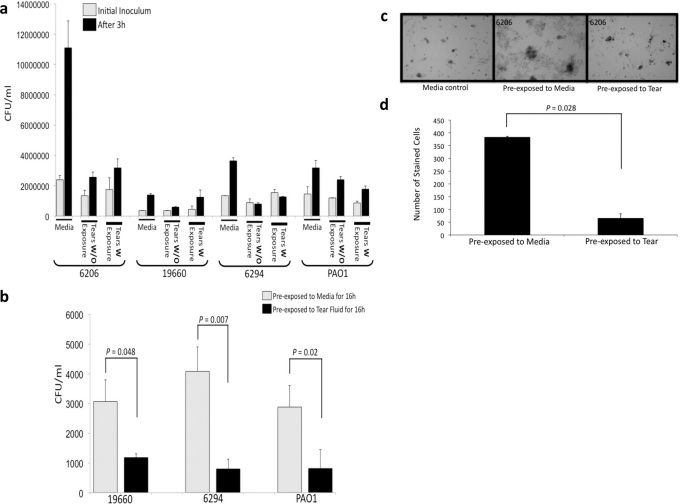
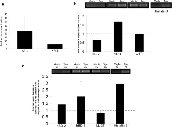
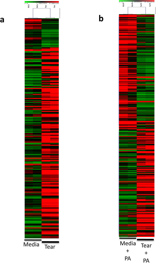
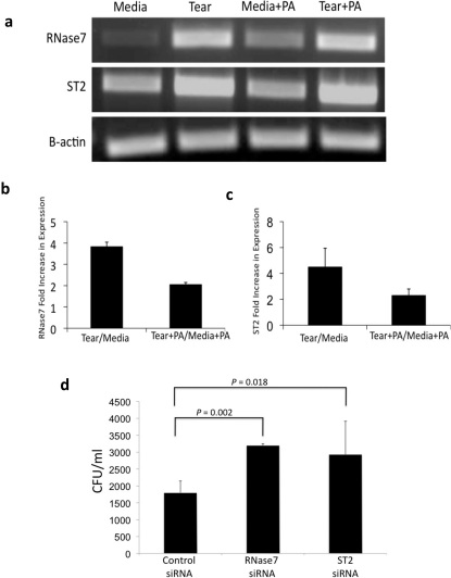
Mucosal epithelial cells, including those at the ocular surface, resist infection by most microbes in vivo but can be susceptible to microbial virulence in vitro. While fluids bathing mucosal surfaces (e.g. tears) contain antimicrobials, potentially pathogenic microbes often thrive in these fluids, suggesting that additional mechanisms mediate epithelial resistance in vivo. Here, tear fluid acted directly upon epithelial cells to enhance their resistance to bacterial invasion and cytotoxicity. Resistance correlated with tear fluid-magnified activation of NFκB and AP-1 transcription factors in epithelial cells in response to bacterial antigens, suggesting priming of innate defense pathways. Further analysis revealed differential regulation of potential epithelial cell defense genes by tears. siRNA knockdown confirmed involvement of at least two factors, RNase7 and ST-2, for which tears increased mRNA levels, in protection against bacterial invasion. Thus, the role of mucosal fluids in defense can include modulation of epithelial immunity, in addition to direct effects on microbes.


Figures

Figure 1
Figure 1. Pre-exposing Epithelial Cells to Human Tear Fluid Protects against P. aeruginosa Invasion and Cytotoxicity, without affecting Tear Bacteriostatic Activity.
a, P. aeruginosa (∼104 cfu) were suspended in cell culture media, tears, or tears pre-exposed to corneal epithelial cells for 3 h at 37 °C. Tear bacteriostatic activity was unaffected by prior exposure to epithelia. b, c, d,. Pre-exposure to tear fluid protected corneal epithelial cells against P. aeruginosa invasion (b) and cytotoxicity (strain 6206) measured by trypan blue staining of dead/dying cells (c, d). Epithelial cells were exposed to tear fluid or cell culture media for 16 h then inoculated with P. aeruginosa (∼104 cfu in cell culture media, 3 h at 37°C, see methods).
Figure 2
Figure 2. Tears Induce AP-1 and NFκB Activation.
a, Corneal epithelial cells were transfected with NFκB or AP-1 transcription factor-responsive firefly luciferase and Renilla luciferase constructs, incubated with tears or cell culture media for 16 h then 3 h with P. aeruginosa antigens. Tears induced a ∼25-fold increase in AP-1 activation, and ∼5-fold increase in NFκB; Fold increase = cells pre-exposed to tears with P. aeruginosa antigens over cells pre-exposed to media with P. aeruginosa antigens b, Histatin-3 and hBD-3 mRNA induction in corneal epithelial cells exposed to tears for 6 h (relative to media control); c, Induction of histatin-3 mRNA in corneal cells pre-exposed to tears (16 h, washed, then inoculated with P. aeruginosa antigens for 3 h).
Figure 3
Figure 3. Microarray Analysis of Epithelial Cells Exposed to Tear and/or P. aeruginosa antigens.
a, b Heat-maps of differentially regulated probe sets in epithelial cells incubated with human tear fluid for 6 h (a) or cells pre-exposed to tear fluid for 16 h followed by 3 h incubation with P. aeruginosa antigens (b). Red and green boxes indicate up- and down-regulation, respectively, of probe sets with tear fluid.
Figure 4
Figure 4. RT-PCR Confirmation of Tear Induced Expression of RNase7 and ST2 and Their Protective Effect against P. aeruginosa Invasion.
a, b, c, Semi-quantitative RT-PCR confirmed that 6 h tear exposure alone or 16 h pre-exposure to tear fluid followed by 3 h incubation with P. aeruginosa antigens induced RNase7 and ST2 mRNA expression in corneal epithelial cells. d, siRNA knockdown of RNase7 or ST2 in corneal epithelial cells increased P. aeruginosa invasion.

Similar articles

Cited by

References

    1. Mun J. J., Tam C., Kowbel D., Hawgood S., Barnett M. J., Evans D. J. & Fleiszig S. M. Clearance of Pseudomonas aeruginosa from a healthy ocular surface involves surfactant protein D and is compromised by bacterial elastase in a murine null-infection model. Infect Immun. 77, 2392–8 (2009). - PMC - PubMed
    1. Fleiszig S. M., Lee E. J., Wu C., Andika R. C., Vallas V., Portoles M. & Frank D. W. Cytotoxic strains of Pseudomonas aeruginosa can damage the intact corneal surface in vitro. CLAO J. 24, 41–7 (1998). - PubMed
    1. Fleiszig S. M., Zaidi T. S. & Pier G. B. Pseudomonas aeruginosa invasion of and multiplication within corneal epithelial cells in vitro. Infect Immun. 63, 4072–7 (1995). - PMC - PubMed
    1. Fleiszig S. M., Kwong M. S. & Evans D. J. Modification of Pseudomonas aeruginosa interactions with corneal epithelial cells by human tear fluid. Infect Immun. 71, 3866–74 (2003). - PMC - PubMed
    1. Kwong M. S., Evans D. J., Ni M., Cowell B. A. & Fleiszig S. M. Human tear fluid protects against Pseudomonas aeruginosa keratitis in a murine experimental model. Infect Immun. 75, 2325–32 (2007). - PMC - PubMed

Publication types