. 2011:1:17.
doi: 10.1038/srep00017. Epub 2011 Jun 17.

Multibudded tubules formed by COPII on artificial liposomes

Affiliations

Multibudded tubules formed by COPII on artificial liposomes

Kirsten Bacia et al. Sci Rep. 2011.

Abstract

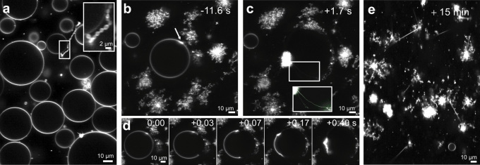
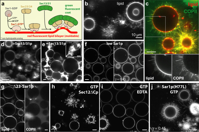
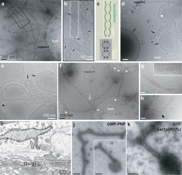
COPII-coated vesicles form at the endoplasmic reticulum for cargo transport to the Golgi apparatus. We used in vitro reconstitution to examine the roles of the COPII scaffold in remodeling the shape of a lipid bilayer. Giant Unilamellar Vesicles were examined using fast confocal fluorescence and cryo-electron microscopy in order to avoid separation steps and minimize mechanical manipulation. COPII showed a preference for high curvature structures, but also sufficient flexibility for binding to low curvatures. The COPII proteins induced beads-on-a-string-like constricted tubules, similar to those previously observed in cells. We speculate about a mechanical pathway for vesicle fission from these multibudded COPII-coated tubules, considering the possibility that withdrawal of the Sar1 amphipathic helix upon GTP hydrolysis leads to lipid bilayer destabilization resulting in fission.


Figures

Figure 1
Figure 1. Membrane tubulation by Sar1p.
Confocal microscopy of fluorescently labeled membranes. (a) Sar1p-GDP control: GUVs remained mostly unaffected. A minority of µm-sized pearling vesicles was observed (inset) [2µM Sar1p, 1µM Sec12ΔCp, 1mM GDP]. (b–d) GUV tubulation by Sar1p-GTP [1µM Sar1p, 0.5µM Sec12ΔCp, 1mM GTPr]. (b) Tubulation starts (arrow). (c) The GUV has exploded into a ‘ball' of thin, wiggling membrane tubules. The inset shows tubules at increased contrast. (d) Timeseries of the explosion. See also Movie S1. (e) Straight, rigid lipid tubules after prolonged incubation with Sar1p-GTP.
Figure 2
Figure 2. Generation of rigid membrane extensions by COPII, visualized by confocal microscopy.
Scale bars = 10 µm. (a) Schematic view. The reaction was reconstituted in vitro by mixing purified red fluorescent GUVs and purified COPII components [2µM Sar1p, 320nM Sec23/24p, 520nM Sec13/31p; 1µM Sec12ΔCp or 2.5mM EDTA (to facilitate nucleotide exchange)]. (b) COPII produced long, rigid membrane extensions that appeared tubular at confocal microscopy resolution. (c) Green fluorescently labeled Sec13/31p showed a preferential COPII localization at highly curved tubules compared to the low curvature GUV. (d/e) Sec13/31p was essential. Extensive rigid tubulation was observed with full COPII and GMP-PNP (even at lowered COPII concentrations: 1µM Sar1p, 160nM Sec23/24p, 260nM Sec13/31p, 2.5mM EDTA, 0.1mM GMP-PNP in panel d). In contrast, in the absence of Sec13/31p, GUVs and smaller liposome material aggregated (panel e). Inset: 3D maximum intensity projection (nonlinear intensity scale). Omitting Sec13/31p from the coat caused GUVs to adhere to each other. See also Fig. S2. (f) At concentrations of only 0.2µM Sar1p [320nM Sec23/24p, 520nM Sec13/31p, 1µM Sec12ΔCp, 1mM GMP-PNP] mostly intact GUVs were observed. Close inspection revealed shorter rigid tubules emanating from the GUVs. Extensions are more easily discernible when using a nonlinear intensity scale (right). (g) The N-terminal amphipathic helix of Sar1p was essential for recruiting COPII. The truncated Δ23 mutant did not recruit COPII marked by a green labeled Sec13/31p (right) to the red labeled lipid bilayer (left) and did not induce tubulation of GUVs [2µM Δ23-Sar1p, 320nM Sec23/24p, 520nM Sec13/31p, 1µM Sec12ΔCp, 1mM GMP-PNP]. (h) With hydrolysable GTP, the COPII proteins produced a different and more heterogeneous picture consisting of intact GUVs, micron-sized vesicles and soft tubules [2µM Sar1p, 320nM Sec23/24p, 520nM Sec13/31p, 1µM Sec12ΔCp, 1mM GTP]. (i) With EDTA instead of Sec12ΔCp, GUVs appeared mostly intact, but some had a granular appearance or were surrounded by micron-sized vesicles [2µM Sar1p, 320nM Sec23/24p, 520nM Sec13/31p, 2.5mM EDTA, 1mM GTP]. (j) In contrast to panels (h) and (i), the Sar1p mutant H77L as part of the COPII coat generated long, straight, rigid extensions with hydrolysable GTP [2µM Sar1p(H77L), 320nM Sec23/24p, 520nM Sec13/31p, 1µM Sec12ΔCp, 1mM GTP].
Figure 3
Figure 3. Generation of multibudded structures by COPII, revealed by EM.
Black scale bars = 200 nm; white = 100 nm. (a–h) Cryo-EM view of membrane extensions generated by COPII [2µM Sar1p, 320nM Sec23/24p, 520nM Sec13/31p, 2.5mM EDTA, 1mM GMP-PNP]. (a) Overview and (b) magnified inset. Tubular extensions were coated (arrows in panel b) and carried constrictions at ≈100 nanometer spacing. (c) Schematic view and model: Drawing of a COPII vesicle string based on cryo-EM. Red denotes the lipid bilayer, green the protein coat. The COPII coat is rigid and may act like an exoskeleton. Bottom: Multibudded vesicles showed elongated shapes. A propensity of the COPII coat to form spheres may contribute to fission at the necks. (d,e) Both large and small vesicles were coated (d,e: arrows), but some large vesicles remained uncoated (e: black arrowhead). An internal vesicle (d: white arrowhead) remained uncoated. (f) Network of multibudded tubules. Occasional branches are seen in (a) and (f) [gray arrow]. Clover-shapes constituted cross-over tubules. Vesicle strings were apparently well-preserved on the carbon support, but often ended at the holes through which the solution had been blotted (white arrowheads). (g) Reconstituted vesicle strings suggest a mechanism for transport of large cargo. Hypothetical cargo is depicted as boxes. A 300 nm long procollagen bundle, for instance, may occupy three consecutive buds, with fission occurring at the adjoining necks. (h) Some vesicle strings were still connected to a larger liposome and appeared similar to ER exit sites in cells. (i) EM of ER exit sites in a wild type fibroblast cell showed multibudded tubules with a striking resemblance to those produced in vitro from artificial liposomes. (j) Multibudded tubules (including occasional branches) in reconstituted samples were also revealed by negative stain EM. The inset shows a multibudded tubule emanating from a collapsed liposome [2µM Sar1p, 320nM Sec23/24p, 520nM Sec13/31p, 2.5mM EDTA, 1mM GMP-PNP]. (k) The use of the GTP-locked Sar1p(H77L) mutant produced multi-budded tubules in the presence of hydrolysable GTP (negative stain; same sample as in Fig. 2j).

References

    1. Futai E., Hamamoto S., Orci L. & Schekman R. GTP/GDP exchange by Sec12p enables COPII vesicle bud formation on synthetic liposomes. EMBO J. 23, 4146–4155 (2004). - PMC - PubMed
    1. Lee M. C. et al. Sar1p N-terminal helix initiates membrane curvature and completes the fission of a COPII vesicle. Cell 122, 605–617 (2005). - PubMed
    1. Saito Y., Kimura K., Oka T. & Nakano A. Activities of mutant Sar1 proteins in guanine nucleotide binding, GTP hydrolysis, and cell-free transport from the endoplasmic reticulum to the Golgi apparatus. J. Biochem. 124, 816–823 (1998). - PubMed
    1. Aridor M., Bannykh S. I., Rowe T. & Balch W. E. Sequential coupling between COPII and COPI vesicle coats in endoplasmic reticulum to Golgi transport. J. Cell Biol. 131, 875–893 (1995). - PMC - PubMed
    1. Bielli A. et al. Regulation of Sar1 NH2 terminus by GTP binding and hydrolysis promotes membrane deformation to control COPII vesicle fission. J. Cell Biol. 171, 919–924 (2005). - PMC - PubMed

Publication types

MeSH terms