. 2011:1:18.
doi: 10.1038/srep00018. Epub 2011 Jun 20.

Biological consequences of nanoscale energy deposition near irradiated heavy atom nanoparticles

Affiliations

Biological consequences of nanoscale energy deposition near irradiated heavy atom nanoparticles

Stephen J McMahon et al. Sci Rep. 2011.

Erratum in

  • Sci Rep. 2013;3:1725

Abstract

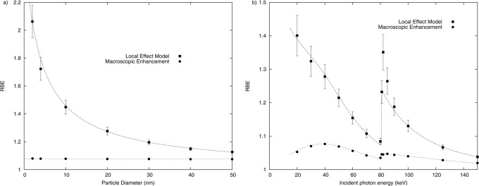
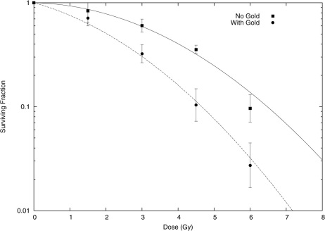
Gold nanoparticles (GNPs) are being proposed as contrast agents to enhance X-ray imaging and radiotherapy, seeking to take advantage of the increased X-ray absorption of gold compared to soft tissue. However, there is a great discrepancy between physically predicted increases in X-ray energy deposition and experimentally observed increases in cell killing. In this work, we present the first calculations which take into account the structure of energy deposition in the nanoscale vicinity of GNPs and relate this to biological outcomes, and show for the first time good agreement with experimentally observed cell killing by the combination of X-rays and GNPs. These results are not only relevant to radiotherapy, but also have implications for applications of heavy atom nanoparticles in biological settings or where human exposure is possible because the localised energy deposition high-lighted by these results may cause complex DNA damage, leading to mutation and carcinogenesis.


Figures

Figure 1
Figure 1. Average energy deposit in the vicinity of a 20 nm gold nanoparticle after a single ionising event by a 40 keV photon.
Energy deposition is here scored in keV in concentric shells around the nanoparticle, broken down into contributions from electrons produced by different processes. In the vicinity of the nanoparticle, Auger electrons produce the dominant contribution, but this falls off rapidly as low-energy electrons are stopped, leaving only the contribution of energetic L-shell Auger electrons beyond 200 nm. Compton electrons are not plotted due to their low number, but are typically roughly 1% of the contribution of the photoelectrons.
Figure 2
Figure 2. Comparison of track structure of ionising events either on the surface (solid lines) or in the bulk (dashed lines) of a 20 nm spherical gold nanoparticle, plotted both in 3D (plot a), and as a 2D projection (plot b).
Here, an incident 50 keV photon (green tracks) interacts with the gold nanoparticle and ejects a number of electrons (red tracks). For the event which occurs in the bulk, the majority of low-energy electrons are stopped immediately in the nanoparticle, allowing only the most energetic and sparsely ionising electrons to escape. By contrast, the surface event also produces a very large shower of low energy electrons who deposit their energy very densely in the vicinity of the nanoparticle, leading to high doses and many ionising events in a small volume.
Figure 3
Figure 3. Probability of different numbers of electrons being emitted from a GNP following an ionisation by a 50 keV photon, as a function of the distance from the ionising event to the nanoparticle surface.
All ionising events in gold typically produce a large number of secondary Auger electrons, but many of these electrons are emitted at low energies and cannot escape because they are stopped in the GNP bulk. At distances a few nanometres within the GNP, a broad distribution of electron yields can be seen, as most Auger electrons can escape following ionisations near the surface. By contrast, at points further from the surface, the distribution is sharply peaked, with only one or two electrons generated following most ionising events.
Figure 4
Figure 4. Average radial doses which are deposited following a single ionising event from 40 keV primary radiation in gold nanoparticles of a variety of sizes.
These doses are calculated by scoring the energy deposited to the water volume in concentric shells around the nanoparticle, and dividing these values by the mass of the water shell. Two features are particularly striking - firstly, areas in the vicinity of the nanoparticle (< 50 nm) see extremely large doses following a typical ionising event. Secondly, small nanoparticles deposit more dose in their local area than larger ones, due to the greater relative contribution from the outer layer of the nanoparticle. The scale and distribution of these doses are broadly similar to those seen in charged particle therapies in the vicinity of a track, which suggests the possible applicability of the local effect model as a way to analyse these results.
Figure 5
Figure 5. Predicted relative biological effectiveness (RBE) of irradiations of MDA-231 cells (survival parameters α = 0.019 ± 0.025, β = 0.052 ± 0.00710) in the presence of 500 µg/mL of gold nanoparticles for a variety of nanoparticle sizes exposed to 40 keV X-rays (a) and 20 nm nanoparticles exposed to a variety of energies (b).
These values were calculated either through as the modification in average dose through the addition of the GNPs (circles), or using the Local Effect Model (squares). The change in macroscopic dose is small, on the order of a few %, in good agreement with the ratio of energy absorption coefficients between gold and water (dotted line). By contrast, the LEM predicts significantly higher effectiveness in all conditions, with strong dependencies on both nanoparticle diameter and incident photon energy. Increasing nanoparticle diameter significantly reduces the RBE, which can be understood as the result of increasing numbers of low-energy electrons being trapped inside the nanoparticle and not contributing to dose in the water volume. The dashed line is an empirical fit, as described in the text. The variation with energy was found to be well described by assuming that each ionising event in a gold nanoparticle contributes a fixed additional probability of a lethal event in a cell, which was characterised by a single empirical fitting parameter, taken to be constant at all energies. Good agreement with modelled values was seen at all points, except immediately above gold's K-edge in plot b, where a significant increase in RBE is observed. This is the result of the majority of the photoelectrons which result from K-shell events being produced at relatively low energies (<5 keV), causing them to contribute much more significantly to short-range dose inhomogeneities than at other energies which are distant from absorption edges.
Figure 6
Figure 6. Experimentally observed cell survival for MDA-231 cells exposed to 160 kVp X-rays with (circles) and without (squares) exposure to 500 µg/mL of 1.9 nm gold nanoparticles, taken from.
In the original analysis, this data was fitted using a conventional linear-quadratic model, to determine if a radiosensitising effect was observed. Here, the data is used to investigate whether the model presented in this work is capable of accurately quantifying the sensitising effects of GNPs. To that end, only the control data was fitted to directly using a linear quadratic, which gave α = 0.019 ± 0.025, β = 0.052 ± 0.007 (solid line). These parameters were used, together with modelled microscopic dose in the vicinity of a 1.9 nm GNP exposed to 160 kVp X-rays, in the Local Effect Model to predict the behaviour of the cells which were exposed to gold nanoparticles, without reference to the experimentally observed results. These theoretical predictions are plotted as the dashed line. The agreement is very good, substantially better than simple energy absorption considerations which predict an increase in damage of just 5% for this gold concentration, which suggests that the microscopic dose in the vicinity of GNPs is a significant contribution to experimentally observed GNP dose enhancement.

References

    1. Webb S. The physical basis of IMRT and inverse planning. Brit. J. Radiol. 76, 678–689 (2003). - PubMed
    1. Hainfeld J. F., Slatkin D. N., Focella T. M. & Smilowitz H. M. Gold nanoparticles: a new X-ray contrast agent. Brit. J. Radiol. 79, 248–53 (2006). - PubMed
    1. Cai Q.-Y. et al. Colloidal gold nanoparticles as a blood-pool contrast agent for X-ray computed tomography in mice. Invest. Radiol. 42, 797–806 (2007). - PubMed
    1. Hainfeld J. F., Dilmanian F. A., Slatkin D. N. & Smilowitz H. M. Radiotherapy enhancement with gold nanoparticles. J. Pharm. Pharmacol. 60, 977–85 (2008). - PubMed
    1. Cho S. Estimation of tumour dose enhancement due to gold nanoparticles during typical radiation treatments: a preliminary Monte Carlo study. Phys. Med. Biol. 50, N163 (2005). - PubMed

Publication types