. 2011:1:47.
doi: 10.1038/srep00047. Epub 2011 Jul 28.

Hyperthermic seizures and aberrant cellular homeostasis in Drosophila dystrophic muscles

Affiliations

Hyperthermic seizures and aberrant cellular homeostasis in Drosophila dystrophic muscles

April K Marrone et al. Sci Rep. 2011.

Abstract

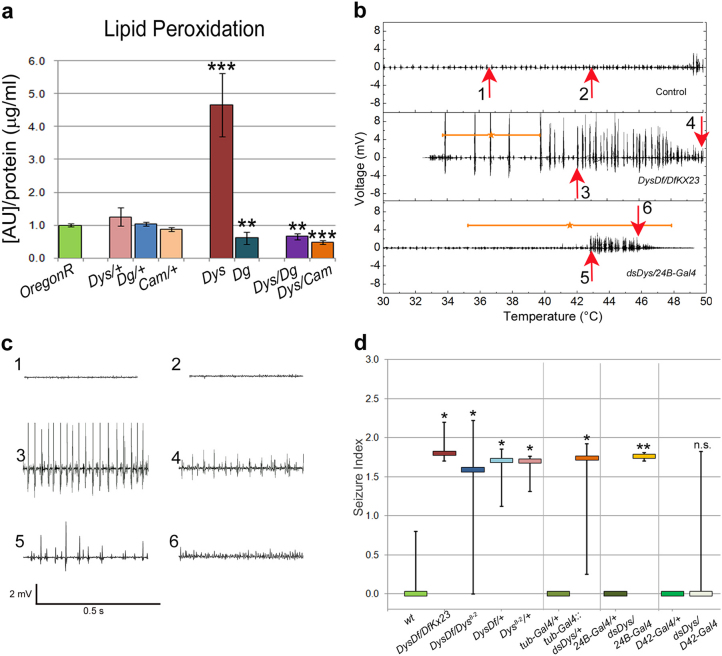
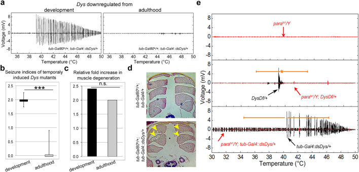
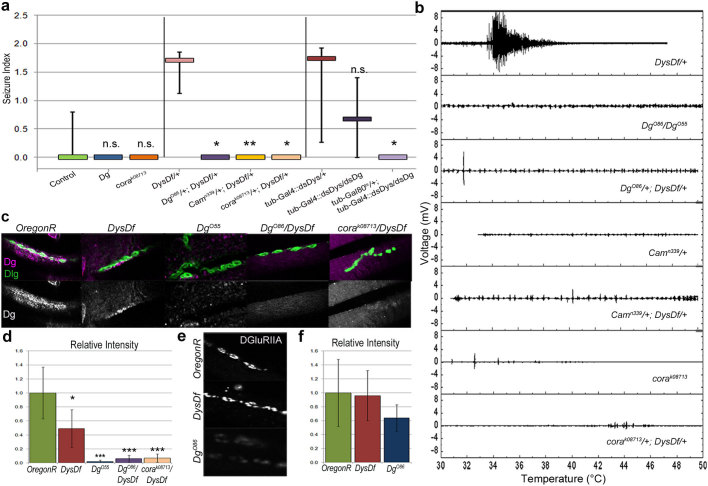
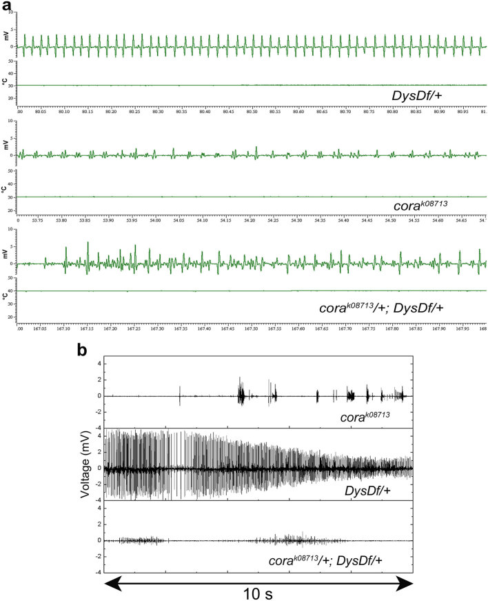
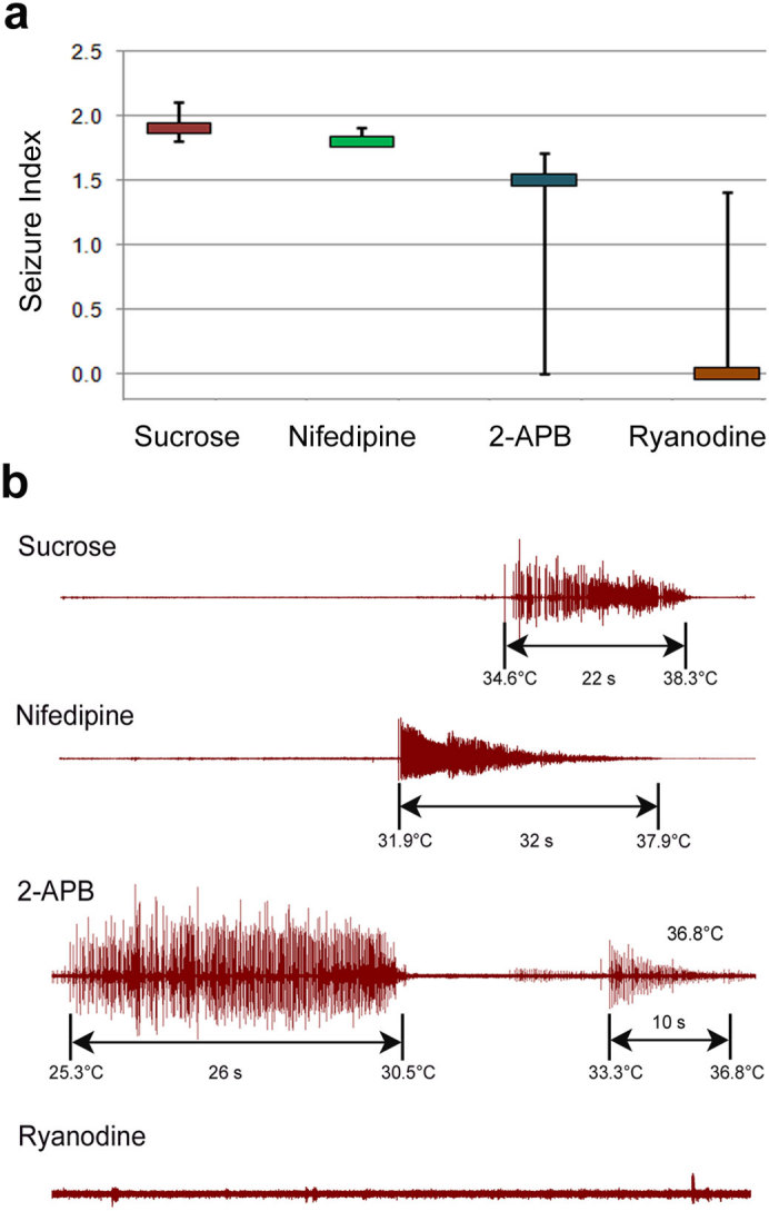
In humans, mutations in the Dystrophin Glycoprotein Complex (DGC) cause muscular dystrophies (MDs) that are associated with muscle loss, seizures and brain abnormalities leading to early death. Using Drosophila as a model to study MD we have found that loss of Dystrophin (Dys) during development leads to heat-sensitive abnormal muscle contractions that are repressed by mutations in Dys's binding partner, Dystroglycan (Dg). Hyperthermic seizures are independent from dystrophic muscle degeneration and rely on neurotransmission, which suggests involvement of the DGC in muscle-neuron communication. Additionally, reduction of the Ca(2+) regulator, Calmodulin or Ca(2+) channel blockage rescues the seizing phenotype, pointing to Ca(2+) mis-regulation in dystrophic muscles. Also, Dys and Dg mutants have antagonistically abnormal cellular levels of ROS, suggesting that the DGC has a function in regulation of muscle cell homeostasis. These data show that muscles deficient for Dys are predisposed to hypercontraction that may result from abnormal neuromuscular junction signaling.


Figures

Figure 1
Figure 1. Dystrophin mutants have elevated ROS and hyperthermic seizures.
(a) Assessment of cellular lipid peroxidation products normalized to protein content in animals housed at 29°C for three days. The axis represents the raw molar absorption after blank subtraction and normalization to protein content. Statistical analysis was done using a one-tailed Student's t-test and the error bars represent the standard deviation. All comparisons are made against OregonR. Exact experimental values and P-values are given in Supplementary Table 1. (b) Electrical output from IFM vs. temperature during seizures of DysDf/DfKX23 and dsDys/24B-Gal4 mutants compared to Control. Stars indicate the average start temperature where the error bar indicates the standard deviation. Arrows indicate where plots in panel c are taken from. (c) Electrical output relative to time where the region examined correlates with the numbered arrows in panel b. Output is displayed over a 0.5 s time period emphasizing the increase in frequency and decrease in amplitude as the seizure progresses. (d) All tested Dys classical and RNAi alleles (levels of Dys mRNA downregulation are shown in Supplementary Table 2) had hyperthermic seizures with similar seizure indices tested in a two-tailed Kruskal-Wallis test where P = 0.316 excluding data from dsDys/D42-Gal4 animals (Supplementary Figure 1, Table 1). Seizure indices of dystrophic mutants are significantly higher when compared to the appropriate control (separated by vertical lines). One to one comparative statistics were done using a one-tailed Mann-Whitney U-test. Error bars indicate the 25th and 75th percentiles of the data spread considering that if an animal does not seize then the index is 0. The Si range of some controls have zero values in the 25th and 75th percentile range, thus it appears that there is no error bar.*P ≤ 0.05, **P ≤ 0.01, ***P ≤ 0.001.
Figure 2
Figure 2. Dystrophic seizures are dependent upon neuronal input that require Dys during development.
(a) Electrical output of animals shifted to the restrictive temperature as late L3 larvae (left) and as adults (right). Dys protein reduction levels using the Gal80 temperature sensitive system are given in Supplementary Table 3 and Supplementary Figure 2. (b) Seizure indices for RNAi mutants after being shifted to the restrictive temperature at different life stages. A Mann-Whitney U-test was used to compare seizure indices upon Dys downregulation in adulthood to Dys downregulation in development (P = 0.007). Dys downregulation in adulthood resulted in a Si that is not significantly different from control (tub-Gal4/+, P = 0.47, Table 1). In addition, animals of the genotype tub-Gal80ts/+;tub-Gal4/+ shifted as adults did not have seizures (n = 2). (c) Bar graphs showing relative fold increase in the frequency of muscle degeneration of Dys RNAi mutants targeting Dys throughout the lifetime or shifted to the restrictive temperature as adult. The percent of muscle degeneration has been normalized to that in control animals under the same conditions (Supplementary Table 4). Statistics for muscle degeneration were done using a one-tailed chi-square test (χ2 = 2.62, P = 0.11). (d) Histological sections of thoracic muscles. Arrows indicate areas of degeneration. (e) The parats1 allele abolishes seizures in DysDf/+ and tub-Gal4::dsDys/+ animals (red plot). *P ≤ 0.05, **P ≤ 0.01, ***P ≤ 0.001.
Figure 3
Figure 3. Loss of Dystroglycan, Coracle or Calmoduline represses the seizure phenotype of Dystrophin mutants.
(a) Seizure indices show that Dg and cora mutants do not have seizures (Table 1) and mutations in Dg, Cam or cora suppress the seizure phenotype in Dys transheterozygous animals (P = 0.032, 0.006 and 0.017 respectively when compared to Dys/+ animals). Animals carrying RNAi constructs against both Dys and Dg also showed a 50% decrease in seizure activity when downregulated during development and 17% when downregulated after development indicating a repression of the phenotype by downregulation of Dg (P = 0.072 and 0.017 respectively compared to Dys RNAi mutant, Table 1). Statistics were done using a one-tailed Mann-Whitney U-test. (b) Electrical output from IFM vs. temperature. (c) In control animals Dg (magenta) is localized to the NMJ. Dys mutants have a decreased amount of Dg at the NMJ, but some Dg localization is still observed. Dg mutants have no Dg localized to the NMJ. Transheterozygous animals (Dg/+;Dys/+, cora/+;Dys/+) show a significant decrease in Dg staining at the NMJ as well. The overall structure and presence of NMJs does not appear to be altered in the mutants as can be seen via Dlg staining (green). (d) In adult abdominal NMJs DGluRIIA is localized similarly in Dys mutants as in the wild type (OregonR) animals; however, Dg loss-of-function mutants have a decreased amount of the receptor. Statistics on relative intensities were done using a Student's t-test compared to OregonR where the error bars represent the average deviation from the mean. *P ≤ 0.05, **P ≤ 0.01, ***P ≤ 0.001.
Figure 4
Figure 4. Cora mutants have a mild seizure phenotype that does not look like and overrides the dystrophic phenotype.
Mutation in the cora gene results in a distinctive electrophysiological phenotype from that seen with Dys heterozygous mutations. (a) corak08713 mutants have electrical output that is not of a consistent frequency. Animals of genotype corak08713/+; DysDf/+ have a phenotype similar to the cora mutant. The output is recorded over 1 second. (b) Examples of the two types of electrical activity measured in corak08713, DysDf/+ and corak08713/+; DysDf/+ animals. The output is recorded over 10 seconds.
Figure 5
Figure 5. Elevated Ca2+ from the SR leads to hyperthermic seizures.
(a) Seizure indices of DysDf mutants after being fed various Ca2+ channel blockers. The control animals were only fed 5% sucrose and the other drugs were given in a 5% sucrose solution. Statistics were calculated using a one-tailed Mann-Whitney U-test where the following P-values were obtained by comparing to sucrose fed animals: Nifedipine: P = 0.421, 2-APB: P = 0.107 and Ryanodine: P = 0.078. (b) Electrical output from dystrophic heated muscles after being fed overnight with sucrose (control), L-type calcium channel blocker (Nifedipine), Ryanodine SR fast calcium channel blocker or IP3R SR slow calcium channel blocker (2-APB). 5–6 animals were measured per condition. Seizure onset and subsiding temperatures are noted as well as seizure duration. Animals fed Nifedipine did not show a decrease in seizure activity. Animals fed 2-APB had additional seizure activity that was occurring spontaneously before heating began that was different in its electrical pattern that would be consistent with the observed phenotype of typical Dys mutants. This is shown here where output occurs for almost 30 s. This is later followed by a seizure at a reasonable temperature that looks like what would be expected for a dystrophic mutant. This extra output was only seen with animals fed this channel blocker, and in general the occurrence of seizures was less than controls (Supplementary Table 5). Animals fed Ryanodine still had seizures 33% of the time, but in general there was a decrease in the seizure indices associated with these animals, therefore we considered this reagent to have a therapeutic effect. *P ≤ 0.05, **P ≤ 0.01, ***P ≤ 0.001.

Similar articles

Cited by

References

    1. Barresi R. & Campbell K. P. Dystroglycan: from biosynthesis to pathogenesis of human disease. J Cell Sci 119, 199–207 (2006). - PubMed
    1. Grady R. M. et al.. Maturation and maintenance of the neuromuscular synapse: genetic evidence for roles of the dystrophin–glycoprotein complex. Neuron 25, 279–293, S0896-6273(00)80894-6 [pii] (2000). - PubMed
    1. Kanagawa M. & Toda T. The genetic and molecular basis of muscular dystrophy: roles of cell-matrix linkage in the pathogenesis. J Hum Genet 51, 915–926, 10.1007/s10038-006-0056-7 (2006). - PubMed
    1. Sciandra F. et al.. Dystroglycan and muscular dystrophies related to the dystrophin-glycoprotein complex. Ann Ist Super Sanita 39, 173–181 (2003). - PubMed
    1. Lim L. E. & Campbell K. P. The sarcoglycan complex in limb-girdle muscular dystrophy. Curr.Opin.Neurol. 11, 443–452 (1998). - PubMed

Publication types