. 2011:1:57.
doi: 10.1038/srep00057. Epub 2011 Aug 8.

Nanofocusing of hard X-ray free electron laser pulses using diamond based Fresnel zone plates

Affiliations

Nanofocusing of hard X-ray free electron laser pulses using diamond based Fresnel zone plates

C David et al. Sci Rep. 2011.

Erratum in

Abstract

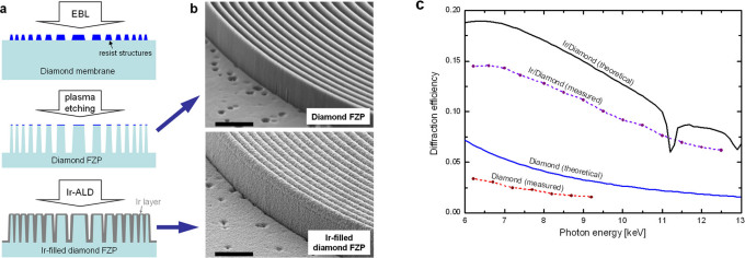
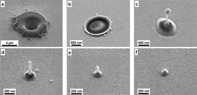
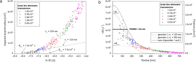
A growing number of X-ray sources based on the free-electron laser (XFEL) principle are presently under construction or have recently started operation. The intense, ultrashort pulses of these sources will enable new insights in many different fields of science. A key problem is to provide x-ray optical elements capable of collecting the largest possible fraction of the radiation and to focus into the smallest possible focus. As a key step towards this goal, we demonstrate here the first nanofocusing of hard XFEL pulses. We developed diamond based Fresnel zone plates capable of withstanding the full beam of the world's most powerful x-ray laser. Using an imprint technique, we measured the focal spot size, which was limited to 320 nm FWHM by the spectral band width of the source. A peak power density in the focal spot of 4×10(17)W/cm(2) was obtained at 70 fs pulse length.


Figures

Figure 1
Figure 1. Gold FZPs damaged by 8 keV LCLS pulses.
SEM images of identical devices with 1 µm high structures and an outermost zone width of 100 nm. (a) no irradiation, (b) after 1,000 pulses, (c) after 10,000 pulses. Pulse power: 1.2 mJ, pulse rate: 60 Hz. The scale bars are 4 µm, the view angle is 45°.
Figure 2
Figure 2. Diamond Fresnel zone plates.
(a) Overview of FZPs etched into a 1.5 mm diameter diamond membrane. The 500 µm diameter of the central FZP is matched to the beam size at the LCLS-XPP end station. (b) Central rings etched 2.1 µm deep. (c) 100 nm wide outermost zones.
Figure 3
Figure 3. Improving the diffraction efficiency of diamond-based Fresnel zone plates.
(a) Overview of the fabrication flow, comprising electron-beam lithography (EBL) to pattern a resist layer, pattern transfer by plasma etching, and filling with Ir by atomic layer deposition (ALD). (b) Comparison of a diamond test FZP with 1.2 µm high structures and 100 nm wide outermost zones before and after filling with Ir by atomic layer deposition. The scale bars are 1 µm. (c) Photon energy dependent diffraction efficiencies of a diamond FZP with 1.8 µm high structures and a Ir-filled diamond FZP with 1.2 µm high structures. The solid lines show theoretical efficiency of FZPs made from these materials with the corresponding structure dimensions.
Figure 4
Figure 4. Imprints created by hard XFEL pulses focused with an Ir-filled diamond FZP.
The Au-coated glass substrates are viewed under an angle of 45°. (a) An unattenuated pulse (∼5×10⁻⁵ J) causes a crater with an inner diameter of 3.5 µm. (b, c) Imprints at ∼5×10⁻⁷ J and 1×10⁻⁷ J result in diameters of 1.1 µm and 690 nm, respectively. (d–f) pulses at ∼2×10⁻⁸ J result in peak fluences close to the ablation threshold of the Au coating. The imprint diameters are 325, 240, and 190 nm. A fountain of molten glass ejected from the substrate and solidified in the center of imprints c–f.
Figure 5
Figure 5. Analysis of the nanofocus spot size of the Ir-filled diamond FZP.
(a) The squared imprint diameters as a function the logarithm of the pulse energy reveals two regions that can be approximated by straight lines corresponding to a double-Gaussian spot with sigma-values of 120 nm and 350 nm, respectively. (b) Beam profile derived from the same data set. The left y-axis shows the inverse pulse energies of the individual shots. The right y-axis is normalised such that the integral energy over the analytical double-Gaussian function is equal to Emax = 5×10⁻⁵ J (see methods).

Similar articles

Cited by

References

    1. Young L. et al.. Femtosecond electronic response of atoms to ultra-intense X-rays. Nature 466, 56–62 (2010). - PubMed
    1. Neutze R., Wouts R., van der Spoel D., Weckert E., and Hajdu J. Potential for biomolecular imaging with femtosecond X-ray pulses. Nature 406, 752–757 (2000). - PubMed
    1. Gaffney K. J., and Chapman H. N. Imaging Atomic Structure and Dynamics with Ultrafast X-ray Scattering. Science 316, 1444–1448 (2007) - PubMed
    1. Chapman H. N. et al.. Femtosecond diffractive imaging with a soft X-ray free electron laser. Nature Phys. 2, 839–843 (2006).
    1. Emma P. et al.. First lasing and operation of an ångstrom-wavelength free-electron laser. Nature Photon. 4, 641–647 (2010).