. 2011:1:76.
doi: 10.1038/srep00076. Epub 2011 Aug 30.

Differential macrophage polarization promotes tissue remodeling and repair in a model of ischemic retinopathy

Affiliations

Differential macrophage polarization promotes tissue remodeling and repair in a model of ischemic retinopathy

Valentina Marchetti et al. Sci Rep. 2011.

Abstract

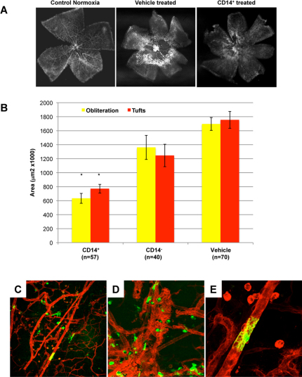
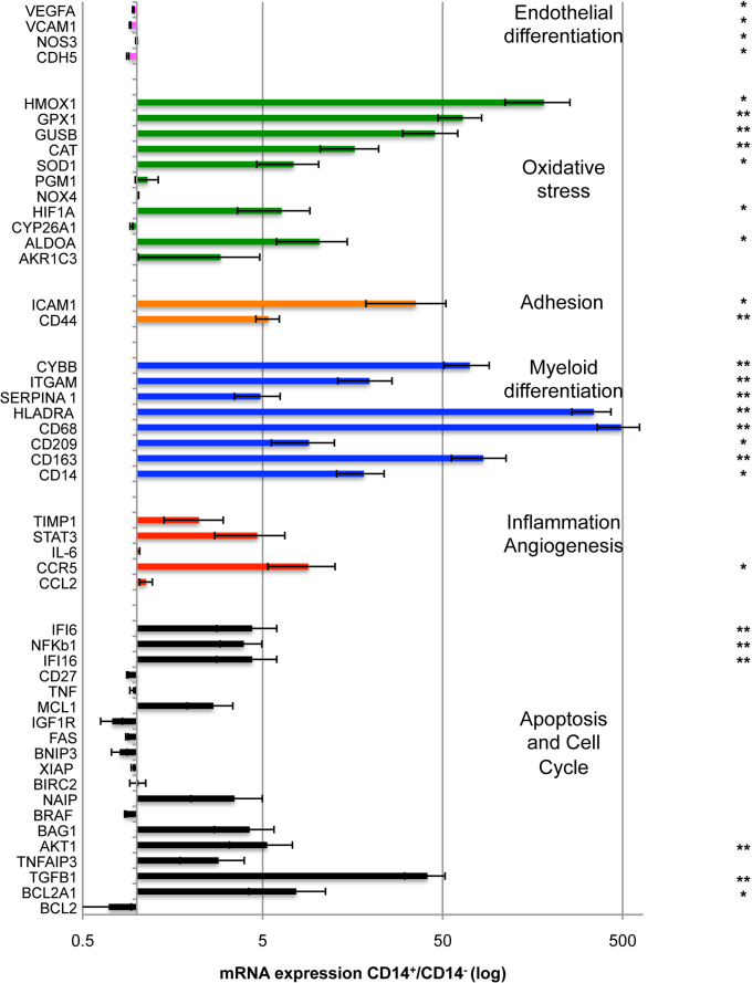
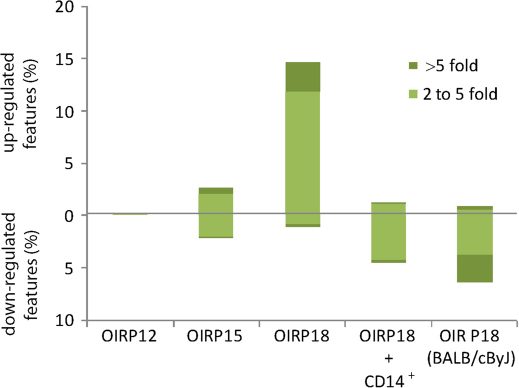
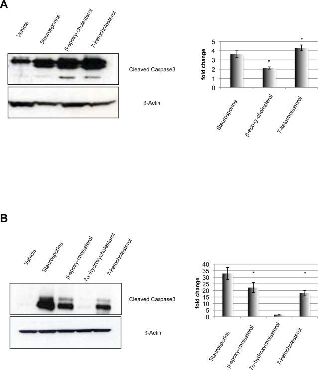
Diabetic retinopathy is the leading cause of visual loss in individuals under the age of 55. Umbilical cord blood (UCB)-derived myeloid progenitor cells have been shown to decrease neuronal damage associated with ischemia in the central nervous system. In this study we show that UCB-derived CD14(+) progenitor cells provide rescue effects in a mouse model of ischemic retinopathy by promoting physiological angiogenesis and reducing associated inflammation. We use confocal microscopy to trace the fate of injected human UCB-derived CD14(+) cells and PCR with species-specific probes to investigate their gene expression profile before and after injection. Metabolomic analysis measures changes induced by CD14(+) cells. Our results demonstrate that human cells differentiate in vivo into M2 macrophages and induce the polarization of resident M2 macrophages. This leads to stabilization of the ischemia-injured retinal vasculature by modulating the inflammatory response, reducing oxidative stress and apoptosis and promoting tissue repair.


Figures

Figure 1
Figure 1. CD14+ cells stabilize and promote normalization of ischemia-injured retinal vasculature in the OIR model.
A) CD14+ cells normalize angiogenesis during hyperoxia and accelerate retinal revascularization. Normal retinas were dissected from C57BL/6J mice and stained with GS lectin at postnatal day 17 (P17). They show a characteristic branching vascular pattern radiating outward from the central optic nerve head (“Control Normoxia”). Exposure to hyperoxia for 5 days (from P7 to P12) leads to central vaso-obliteration. Once the mice return to normoxia, neovascularisation occurs at the interface between perfused peripheral, and non-perfused central, retina. Treatment with vehicle (“Vehicle treated”) at P7 does not alter the vaso-obliteration or neovascularisation. In contrast, treatment at P7 with hUCB-derived CD14+ cells leads to normalization of the retinal vasculature (“CD14+ treated”). B) Quantification of retinas treated with hUCB-derived cell populations. Retinas were analyzed at P17 using GS-lectin staining for retinal vessel obliteration (yellow bars) and tuft formation (neovascularization) (red bars) in retinal whole mounts. No significant difference was observed between vehicle and CD14 cells when injected intravitreally at P7. Obliteration and neovascularization are reduced by 63% and 56% respectively, compared to vehicle-treated retinas (n = 57, n = 40, n = 70, n = number of eyes) (*P <0.001 Bonferroni corrected t-test). C) D) And E) Ad5F16-eGFP hUCB-derived CD14+ cells (green) target and differentiate along the mouse retina vasculature (red) at P17 (10x, 20x and 40X respectively).
Figure 2
Figure 2. CD14+ cells promote angiogenesis and recruit endogenous proangiogenic cells in hypoxic areas (A) At P12 the obliteration area in CD14+ treated eyes is reduced by 50% compared to vehicle-treated retinas (n = 8, n = number of eyes for each condition).
(B) Lectin-positive cells in areas of vaso-obliteration are increased 2.2- fold after intravitreal injection of CD14+ cells relative to vehicle injection (n = 6, n = number of eyes for each condition) (*P <0.001 Bonferroni corrected t-test). (C) 10x confocal images of retinas treated with CD14+ shows high recruitment of lectin-positive cells with microglial-like morphology. (D) Lectin-positive cells are absent in vehicle-injected retinas.
Figure 3
Figure 3. Human UCB-derived CD14+ cells regulate oxidative stress and apoptosis, and express markers of myeloid cell differentiation in the OIR model.
Several human genes are up-regulated at P17 in the CD14+-treated retinas compared to retinas injected with the negative fraction, CD14cells and compared to retinas injected with the vehicle, DPBS. A fold increase of 1.6 or greater of anti-apoptotic genes is observed (black) (IFI6, IFI16, AKT1, BCL2A1). Human anti-oxidative stress genes are significantly up regulated when CD14+ cells are injected compared to the negative fraction (green). Numerous genes characteristic of myeloid cell differentiation are highly expressed in the CD14+-treated retinas (blue). The CD14+ cells increase expression of vascular adhesion molecules (ICAM1) and the extracellular matrix receptor (CD44) (orange). Genes characteristic of endothelial cell differentiation (VEGF) and inflammation (TNF, IL6) are not significantly different between the CD14+ cells and the retinas treated with the negative fraction. (n = 12, n = number of retinas independently extracted and analyzed for each treatment) (*P <0.01; **P <0.001).
Figure 4
Figure 4. Global metabolomic analysis of the OIR model.
Bars indicate the percentage of up-regulated or down-regulated mass spectrometry features (*P <0.01) in the OIR retinas relative to normoxia at post natal day 12, 15 and 18. Results correspond to C57BL/6J mouse strain unless otherwise stated. 8–12 independently extracted retinal tissues were analyzed for each time point or treatment, n = 8–12).
Figure 5
Figure 5. Oxysterols induce apoptosis in endothelial cells and astrocytes in vitro.
Western Blot analysis shows the cleavage of Caspase-3 induced by β−epoxycholesterol and 7-ketocholesterol in Bovine Aortic Endothelial cells (A) and C8-D1A astrocytes (B). Staurosporin (ST) was used as positive control. Actin level is reported to normalize the quantity of protein in each sample (n = 3, n = number of experiments) (Bonferroni corrected t-test: *P <0.01).
Figure 6
Figure 6. Oxysterols are toxic for astrocytes and muller glia cells in vivo.
Immunohistochemical analysis using lectin (red, blood vessels) and antibody to GFAP (green, astrocytes and activated Muller glia cells) demonstrates the normal retina (A) or abnormalities observed with degeneration of astrocytes in the inner retina after intravitreal injection of β-epoxy-cholesterol (B) or 7-ketocholesterol (C). Insets in A, B and C represent 20X magnification views of GFAP expression. Inset figures a, b, c show 20X magnification views of whole mount retinas from the same specimen; astrocytes (green) appear damaged and the muller glia cells (green dots) are visible in the superficial plexus of the vasculature (red) demonstrating severe gliosis (n = 5, n = number of eyes for each treatment). HUCB derived CD14+ cells prevent Muller Glia cells activation in OIR retinas. Immunohistochemical analysis using an antibody to GFAP demonstrates degeneration of astrocytes layer and gliosis (red arrows) in P17 OIR retinas (D). Treatment with HUCB CD14+ cells prevents this damage (E) (n = 6, n = number eyes for each condition).
Figure 7
Figure 7. Intravitreal injection of M2 macrophages derived from CD14+ cells reduces oxygen induced neovascularization.
CD14+ cells were differentiated into M1 or M2 cells and injected into the vitreous of P7 mice just prior to high oxygen exposure in the OIR model. At P17 the areas of obliteration (yellow) and neovascular tuft formation (red) were quantified as described in the experimental procedures. Both M1 and M2 populations significantly reduce the areas of obliteration compared to the control-treated eyes (n = 17, 15, 10 and 10 respectively, n = number of eyes for each treatment) (Bonferroni corrected t-test, *P <0.001). M2 cells are significantly more effective than the M1 cells at reducing the area of neovascularisation compared to the vehicle- or non-injected eyes. (B) At P17, CD14+-treated retinas show recruitment of mouse macrophages expressing mannose receptor (MR) (green). GS-lectin staining shows mouse vasculature (red). (C) MR positive cells are not observed in vehicle-treated eyes (20X).

References

    1. Newcomb J. D., Sanberg P. R., Klasko S. K. & Willing A. E. Umbilical cord blood research: current and future perspectives. Cell Transplant 16, 151–158 (2007). - PMC - PubMed
    1. Broxmeyer H. E. et al.. Growth characteristics and expansion of human umbilical cord blood and estimation of its potential for transplantation in adults. Proc Natl Acad Sci U S A 89, 4109–4113 (1992). - PMC - PubMed
    1. Roncarolo M. G., Bigler M., Ciuti E., Martino S. & Tovo P. A. Immune responses by cord blood cells. Blood Cells 20, 573–585; discussion 585–576 (1994). - PubMed
    1. Henning R. J. et al.. Human cord blood cells and myocardial infarction: effect of dose and route of administration on infarct size. Cell Transplant 16, 907–917 (2007). - PubMed
    1. Womble T. A., Green S., Sanberg P. R., Pennypacker K. R. & Willing A. E. CD14(+) human umbilical cord blood cells are essential for neurological recovery following MCAO. Cell Transplantation 17, 485–486 (2008).

Publication types

Substances