

Bringing the real world into the fMRI scanner: repetition effects for pictures versus real objects
- PMID: 22355647
- PMCID: PMC3216611
- DOI: 10.1038/srep00130
Bringing the real world into the fMRI scanner: repetition effects for pictures versus real objects
Abstract
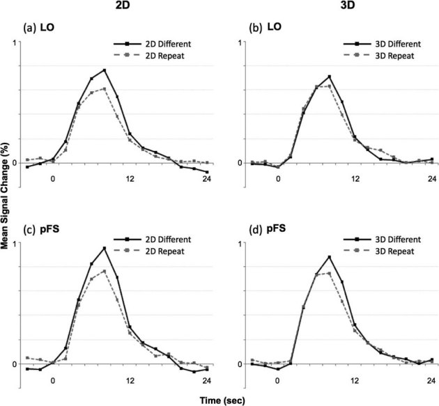
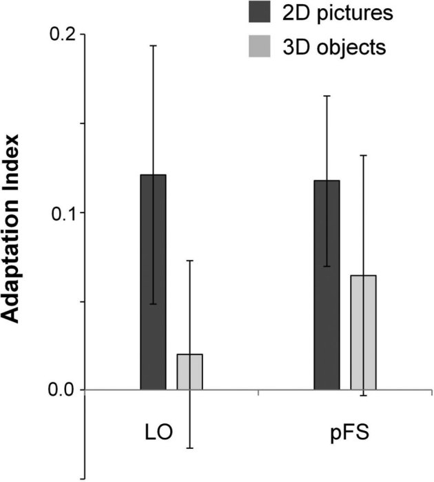
Our understanding of the neural underpinnings of perception is largely built upon studies employing 2-dimensional (2D) planar images. Here we used slow event-related functional imaging in humans to examine whether neural populations show a characteristic repetition-related change in haemodynamic response for real-world 3-dimensional (3D) objects, an effect commonly observed using 2D images. As expected, trials involving 2D pictures of objects produced robust repetition effects within classic object-selective cortical regions along the ventral and dorsal visual processing streams. Surprisingly, however, repetition effects were weak, if not absent on trials involving the 3D objects. These results suggest that the neural mechanisms involved in processing real objects may therefore be distinct from those that arise when we encounter a 2D representation of the same items. These preliminary results suggest the need for further research with ecologically valid stimuli in other imaging designs to broaden our understanding of the neural mechanisms underlying human vision.
Figures

Similar articles
-
Interaction between Scene and Object Processing Revealed by Human fMRI and MEG Decoding.J Neurosci. 2017 Aug 9;37(32):7700-7710. doi: 10.1523/JNEUROSCI.0582-17.2017. Epub 2017 Jul 7. J Neurosci. 2017. PMID: 28687603 Free PMC article.
-
Selective attention modulates neural substrates of repetition priming and "implicit" visual memory: suppressions and enhancements revealed by FMRI.J Cogn Neurosci. 2005 Aug;17(8):1245-60. doi: 10.1162/0898929055002409. J Cogn Neurosci. 2005. PMID: 16197681 Clinical Trial.
-
Cortical responses to invisible objects in the human dorsal and ventral pathways.Nat Neurosci. 2005 Oct;8(10):1380-5. doi: 10.1038/nn1537. Epub 2005 Sep 4. Nat Neurosci. 2005. PMID: 16136038
-
Appearance isn't everything: news on object representation in cortex.Neuron. 2007 Aug 2;55(3):341-4. doi: 10.1016/j.neuron.2007.07.017. Neuron. 2007. PMID: 17678848 Review.
-
Human cortical areas underlying the perception of optic flow: brain imaging studies.Int Rev Neurobiol. 2000;44:269-92. doi: 10.1016/s0074-7742(08)60746-1. Int Rev Neurobiol. 2000. PMID: 10605650 Review.
Cited by
-
Identifying Cortical Substrates Underlying the Phenomenology of Stereopsis and Realness: A Pilot fMRI Study.Front Neurosci. 2019 Jul 11;13:646. doi: 10.3389/fnins.2019.00646. eCollection 2019. Front Neurosci. 2019. PMID: 31354404 Free PMC article.
-
Grasping performance depends upon the richness of hand feedback.Exp Brain Res. 2021 Mar;239(3):835-846. doi: 10.1007/s00221-020-06025-0. Epub 2021 Jan 5. Exp Brain Res. 2021. PMID: 33403432
-
Disentangling Representations of Object and Grasp Properties in the Human Brain.J Neurosci. 2016 Jul 20;36(29):7648-62. doi: 10.1523/JNEUROSCI.0313-16.2016. J Neurosci. 2016. PMID: 27445143 Free PMC article.
-
The Dorsal Visual Pathway Represents Object-Centered Spatial Relations for Object Recognition.J Neurosci. 2022 Jun 8;42(23):4693-4710. doi: 10.1523/JNEUROSCI.2257-21.2022. Epub 2022 May 4. J Neurosci. 2022. PMID: 35508386 Free PMC article.
-
The Spatiotemporal Neural Dynamics of Object Recognition for Natural Images and Line Drawings.J Neurosci. 2023 Jan 18;43(3):484-500. doi: 10.1523/JNEUROSCI.1546-22.2022. Epub 2022 Dec 19. J Neurosci. 2023. PMID: 36535769 Free PMC article.
References
-
- Kanwisher N. G., Chun M. M., McDermott J. & Ledden P. J. Functional imaging of human visual recognition. Cogn. Brain Res. 5, 55–67 (1996). - PubMed
-
- Grill-Spector K. The neural basis of object perception. Curr. Opin. Neurobiol. 13, 159–166 (2003). - PubMed
-
- Konen C. S. & Kastner S. Two hierarchically organized neural systems for object information in human visual cortex. Nat. Neurosci. 11, 224–231 (2008). - PubMed
Publication types
MeSH terms
Substances
Grants and funding
LinkOut - more resources
Full Text Sources
Other Literature Sources
Medical
Research Materials
