. 2011:1:140.
doi: 10.1038/srep00140. Epub 2011 Nov 3.

Huntingtin mediates dendritic transport of β-actin mRNA in rat neurons

Affiliations

Huntingtin mediates dendritic transport of β-actin mRNA in rat neurons

Bin Ma et al. Sci Rep. 2011.

Abstract

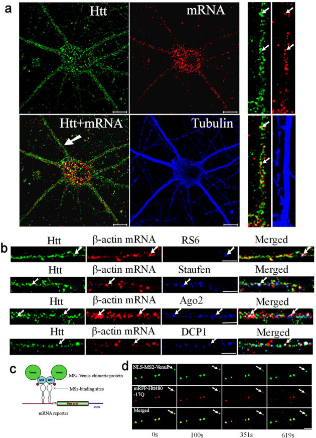
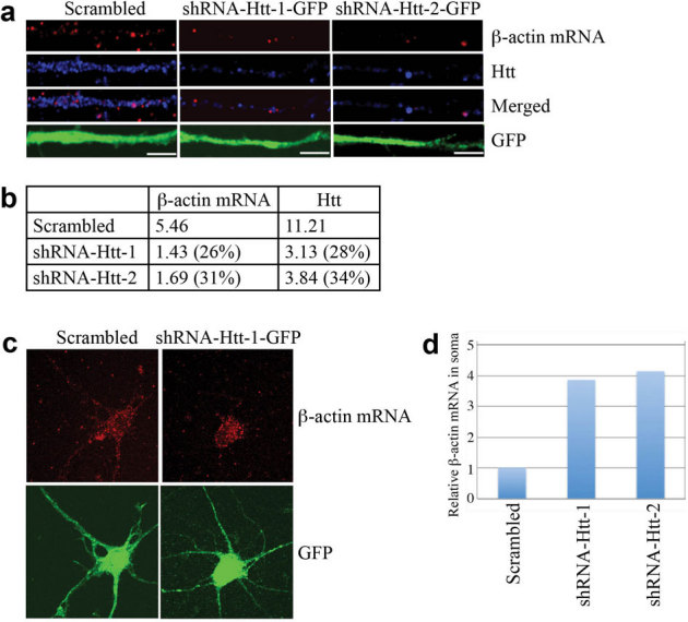
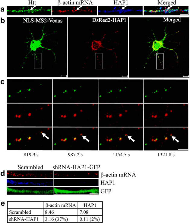
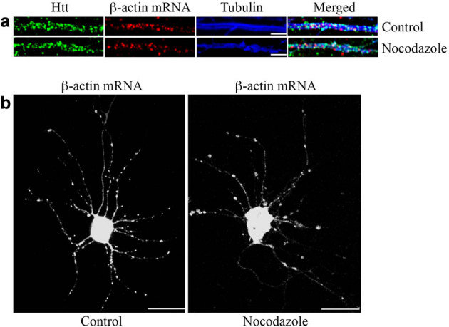
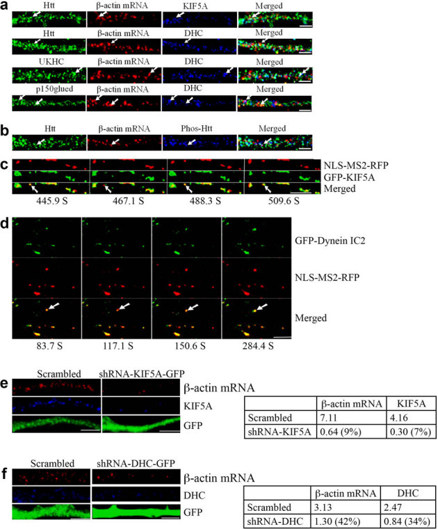
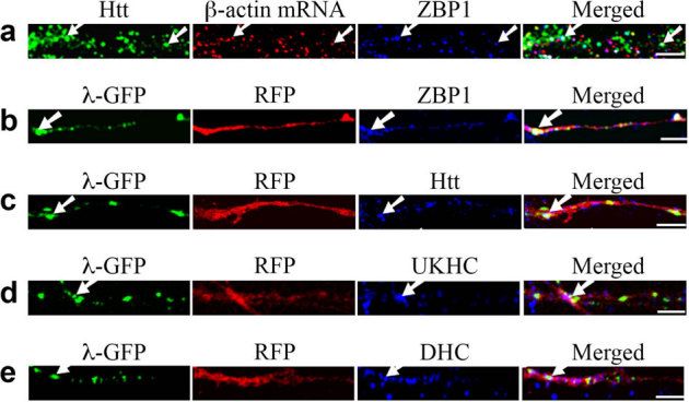
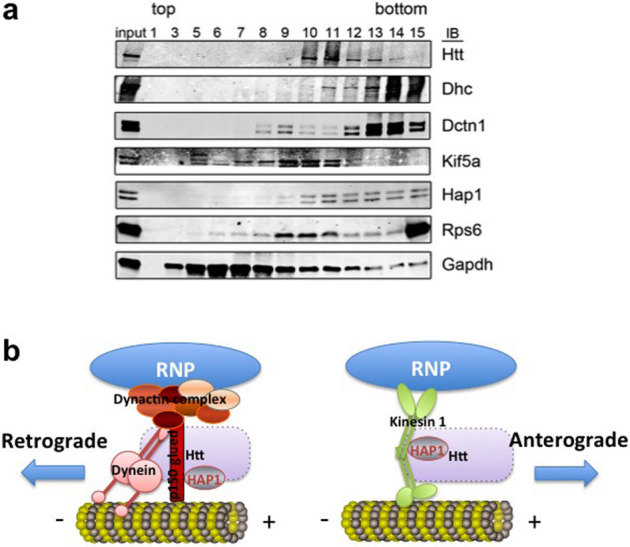
Transport of mRNAs to diverse neuronal locations via RNA granules serves an important function in regulating protein synthesis within restricted sub-cellular domains. We recently detected the Huntington's disease protein huntingtin (Htt) in dendritic RNA granules; however, the functional significance of this localization is not known. Here we report that Htt and the huntingtin-associated protein 1 (HAP1) are co-localized with the microtubule motor proteins, the KIF5A kinesin and dynein, during dendritic transport of β-actin mRNA. Live cell imaging demonstrated that β-actin mRNA is associated with Htt, HAP1, and dynein intermediate chain in cultured neurons. Reduction in the levels of Htt, HAP1, KIF5A, and dynein heavy chain by lentiviral-based shRNAs resulted in a reduction in the transport of β-actin mRNA. These findings support a role for Htt in participating in the mRNA transport machinery that also contains HAP1, KIF5A, and dynein.


Figures

Figure 1
Figure 1. Htt is involved in the dendritic transport of β-actin mRNA in rat cortical neurons.
(a) Co-localization of Htt and β-actin mRNA in cortical neurons (DIV 9). Htt, β-actin mRNA, and α-tubulin are shown in green, red, and blue, respectively. The panels at right are high-magnification views of a single dendrite pointed with a white arrow in the merged image. The arrows indicate the co-localization of Htt and mRNA. Scale bar: 10.0 μm. (b) Htt, β-actin mRNA, and indicated RNP-associated protein (RPS6, staufen, Ago2, DCP1) are shown in green, red, and blue, respectively. Co-localization of Htt/mRNA, Htt/RNP-associated protein, and mRNA/RNP-associated protein is shown in yellow, cyan, and magenta, respectively, in the merged panel. The arrows indicate the co-localization of Htt, mRNA, and RNP-associated protein (appear as white). The left part of each image is the proximal part of the dendrite. Scale bar: 5.0 μm. (c) Schematic representation of the MS2-Venus reporter system for the visualization of β-actin mRNA in live cortical neurons. (d) Co-trafficking of Htt with β-actin mRNA in rat cortical neurons. β-actin mRNA is shown in green and co-transfected mRFP-Htt480-17Q in red. Four cropped images from a time-lapse series captured over 652.5 seconds are shown. The left part of each image is the proximal part of the dendrite. The arrows indicate one retrogradely moving RNA granule with Htt in the neuron. The distance that the granule traveled was 2.78 μm. Scale bar: 5.0 μm.
Figure 2
Figure 2. Knockdown of Htt decreases β-actin mRNA levels in rat neurons leading to accumulation in the soma.
(a) DIV 4 cortical neurons were infected with lentivirus expressing scrambled shRNA, shRNA-Htt-1, or shRNA-Htt-2, and probed at DIV 8 for β-actin mRNA (red), Htt (blue), GFP (green, with anti-GFP antibody). Representative images from each experiment are shown. The left part of each image is the proximal part of the dendrite. Scale bar: 5.0 μm. (b) Relative intensity of Htt and β-actin mRNA shown in (a), normalized to the area of GFP staining, which represents size of dendrite. The percent shown in parentheses indicates extent of knockdown for Htt, and reduction in signal for β-actin mRNA relative to scrambled. (c) Representative images of β-actin mRNA and GFP in a whole neuron after knockdown of Htt. Scale bar: 10.0 μm. (d) Quantitative analysis of enrichment of β-actin mRNA in the soma shown in (c). Intensity of β-actin mRNA in the soma was divided by total β-actin mRNA. Control sample (infected with lentivirus carrying scrambled shRNA) was set to 1. n = 2.
Figure 3
Figure 3. HAP1 is involved in the dendritic targeting of β-actin mRNA.
(a) Co-localization of Htt, β-actin mRNA, and HAP1 in rat cortical neurons (DIV 9). Htt, β-actin mRNA, and HAP1 are shown in green, red, and blue, respectively. The left part of each image is the proximal part of the dendrite. The arrows indicate the co-localization of Htt, β-actin mRNA, and HAP1. Scale bar: 5.0 μm. (b) Co-trafficking of HAP1 with β-actin mRNA in rat cortical neurons (DIV 6). β-actin mRNA is seen in green and HAP1 in red. HAP1 co-localizing with β-actin mRNA is in yellow in the merged panel. Scale bar: 10.0 μm. Part of the boxed area is shown at a high-magnification in (c). (c) Four cropped images from a time-lapse series captured over 1656.5 seconds. The left part of each image is the proximal part of the dendrite. The arrows indicate retrograde movement of one mRNA granule with HAP1 in the neuron. The distance that the granule traveled was 4.03 μm. Scale bar: 5.0 μm. (d) Knockdown of HAP1 decreases β-actin mRNA levels in rat neurons. DIV 4 cortical neurons were infected with lentivirus expressing scrambled shRNA or shRNA-HAP1 and probed at DIV 8 for β-actin mRNA (red), HAP1 (blue), and GFP (green). The left part of each image is the proximal part of the dendrite. Scale bar: 5.0 μm. (e) Quantitative analysis of the intensity of β-actin mRNA and HAP1 normalized to GFP. The percent shown in parentheses indicates extent of knockdown for HAP1, and reduction in signal for β-actin mRNA relative to scrambled.
Figure 4
Figure 4. Transport of mRNA in dendrites is microtubule-dependent.
(a) The distribution of β-actin mRNA in the dendrites of DIV 9 control neuron and neuron treated with nocodazole. In the control neuron, β-actin mRNA is associated with microtubules. The staining of mRNAs does not change significantly after a 30-minute treatment with 2 μg/ml nocodazole. Scale bar: 5.0 μm. (b) Visualization of β-actin mRNA in the control neuron and neuron after nocodazole treatment (2 μg/ml, 30 minutes). Scale bar: 10.0 μm.
Figure 5
Figure 5. Motor proteins kinesin-1 and dynein are involved in the dendritic transport of β-actin mRNA.
The left part of each image is the proximal part of the dendrite. DHC: dynein heavy chain. Scale bar: 5.0 μm. (a) KIF5A and dynein co-localize with β-actin mRNA in rat cortical neurons (DIV 9). The arrows indicate the co-localization of three targets. UKHC: kinesin-1 heavy chain. (b) Co-localization of Htt, β-actin mRNA, and Phos-Htt in rat cortical neurons (DIV 9). (c) Live cell co-trafficking of KIF5A with β-actin mRNA in rat cortical neurons was imaged over 509.6 seconds. β-actin mRNA (detected by NLS-MS2-RFP) is in red and KIF5A (GFP) in green. The arrow indicates an mRNA granule that moves anterogradely along the dendrite. The distance that the granule traveled was 1.39 μm. The velocity of the granule is 0.013 μm/s. (d) Co-trafficking of DIC with β-actin mRNA in rat cortical neurons was imaged live over 1040.4 seconds. β-actin mRNA (detected by NLS-MS2-RFP) is in red and DIC (EGFP) in green. The arrow indicates an mRNA granule that moves retrogradely along dendrites. The distance that the granule traveled was 1.57 μm. DIC: dynein intermediate chain. (e–f) Knockdown of KIF5A or DHC decreases β-actin mRNA levels in rat neurons. DIV 4 cortical neurons were infected with a lentivirus expressing scrambled shRNA, shRNA-KIF5A, or shRNA-DHC and probed at DIV 8 for β-actin mRNA (red), KIF5A or DHC (blue), and GFP (green). The left part of each image is the proximal part of the dendrite. The right panel shows quantitative analysis of the normalized intensity. The percent shown in parentheses indicates extent of knockdown for KIF5A or DHC, and reduction in signal for β-actin mRNA relative to scrambled.
Figure 6
Figure 6. The zipcode sequence in the 3′UTR of β-actin mRNA is sufficient for dendritic targeting and co-localization with Htt in rat neurons.
The left part of each image is the proximal part of the dendrite. Scale bar: 5.0 μm. (a) cortical neurons (DIV 9) were probed for Htt (green), β-actin mRNA (red), and ZBP1 (blue). The arrows indicate the co-localization of Htt, β-actin mRNA, and ZBP1. (b–e) β-actin-zipcode mRNA is visualized by co-transfection of RFP-4xboxB-β-actin-zipcode reporter and λN-GFP, which binds to the 4xboxB sequence. RFP and mRNA are shown in red and green, respectively. Co-localization of mRNA (green) and endogenous proteins (blue) of the transport machinery is indicated by arrows. Co-localization of the translated RFP and endogenous ZBP1, Htt, kinesin-1 (UKHC), and dynein HC are shown in (b), (c), (d), and (e), respectively. Although λN-GFP also represents RFP mRNA, translated RFP diffuses within the dendrite resulting in the loss of punctate pattern of mRNA. DHC: dynein heavy chain.
Figure 7
Figure 7. Htt co-fractionates with microtubule motors and Rps6 in mouse brain.
(a) Western blot of glycerol gradient fractions for indicated proteins (at right). Increasing numbers correspond to lower (denser) position in the gradient. The material loaded onto the gradient is represented in the input lane (3%). Dhc, dynein heavy chain. Dctn 1, dynactin 1. (b) Proposed model of Htt-mediated RNP transport in the dendrite. Htt/HAP1/dynein/dynactin complex is involved in retrograde transport of RNP, whereas Htt/HAP1/kinesin is involved in anterograde transport of RNP. Exact placement of Htt is not known (indicated by a dashed rectangle).

Similar articles

Cited by

References

    1. Borrell-Pages M., Zala D., Humbert S. & Saudou F. Huntington's disease: from huntingtin function and dysfunction to therapeutic strategies. Cell Mol Life Sci 63, 2642–2660 (2006). - PMC - PubMed
    1. Caviston J. P. & Holzbaur E. L. Huntingtin as an essential integrator of intracellular vesicular trafficking. Trends Cell Biol 19, 147–155 (2009). - PMC - PubMed
    1. Gauthier L. R. et al.. Huntingtin controls neurotrophic support and survival of neurons by enhancing BDNF vesicular transport along microtubules. Cell 118, 127–138 (2004). - PubMed
    1. Gunawardena S. et al.. Disruption of axonal transport by loss of huntingtin or expression of pathogenic polyQ proteins in Drosophila. Neuron 40, 25–40 (2003). - PubMed
    1. Bramham C. R. & Wells D. G. Dendritic mRNA: transport, translation and function. Nat Rev Neurosci 8, 776–789 (2007). - PubMed

Publication types

MeSH terms