

Dynein light chain 1 functions in somatic cyst cells regulate spermatogonial divisions in Drosophila
- PMID: 22355688
- PMCID: PMC3240984
- DOI: 10.1038/srep00173
Dynein light chain 1 functions in somatic cyst cells regulate spermatogonial divisions in Drosophila
Abstract
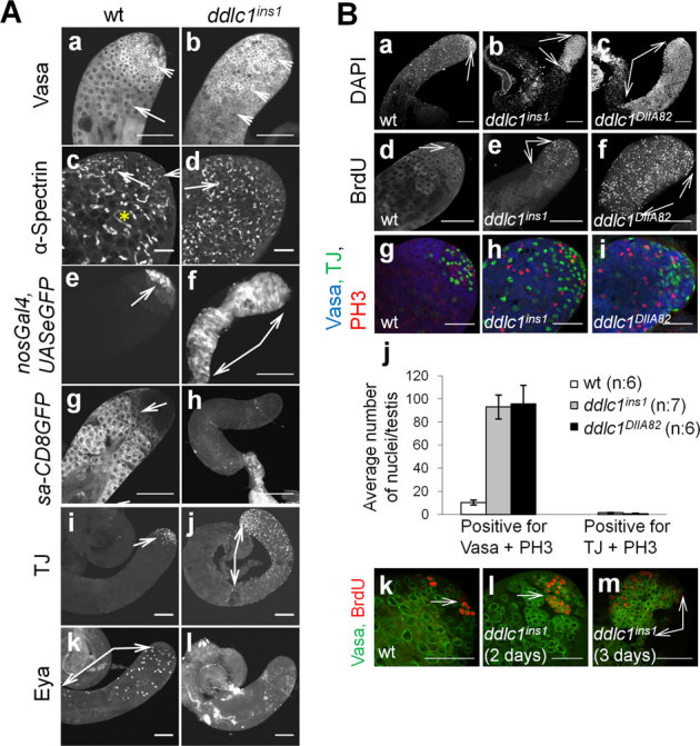
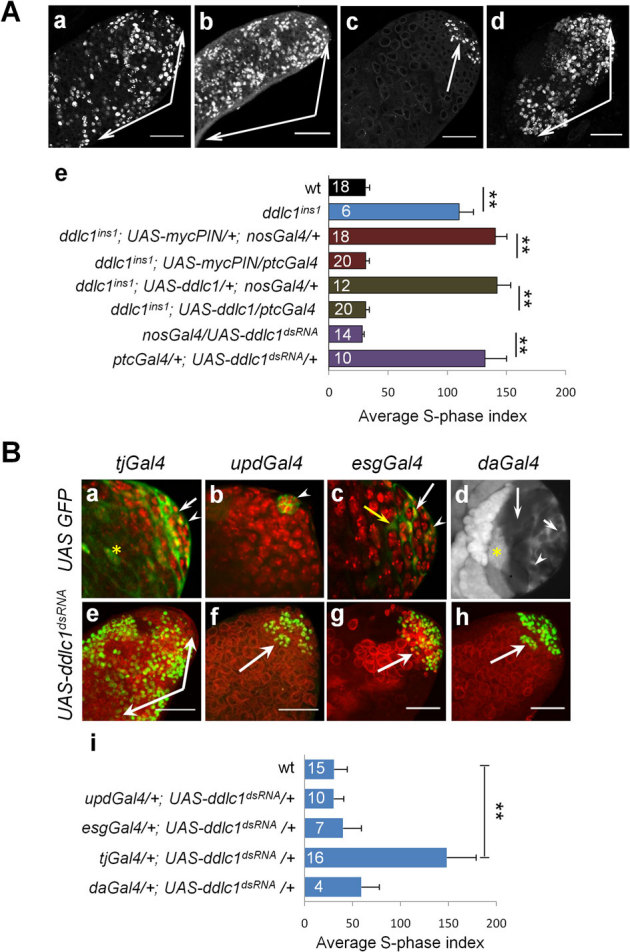
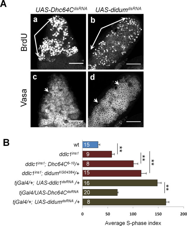
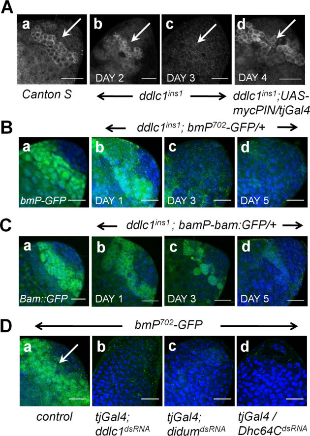
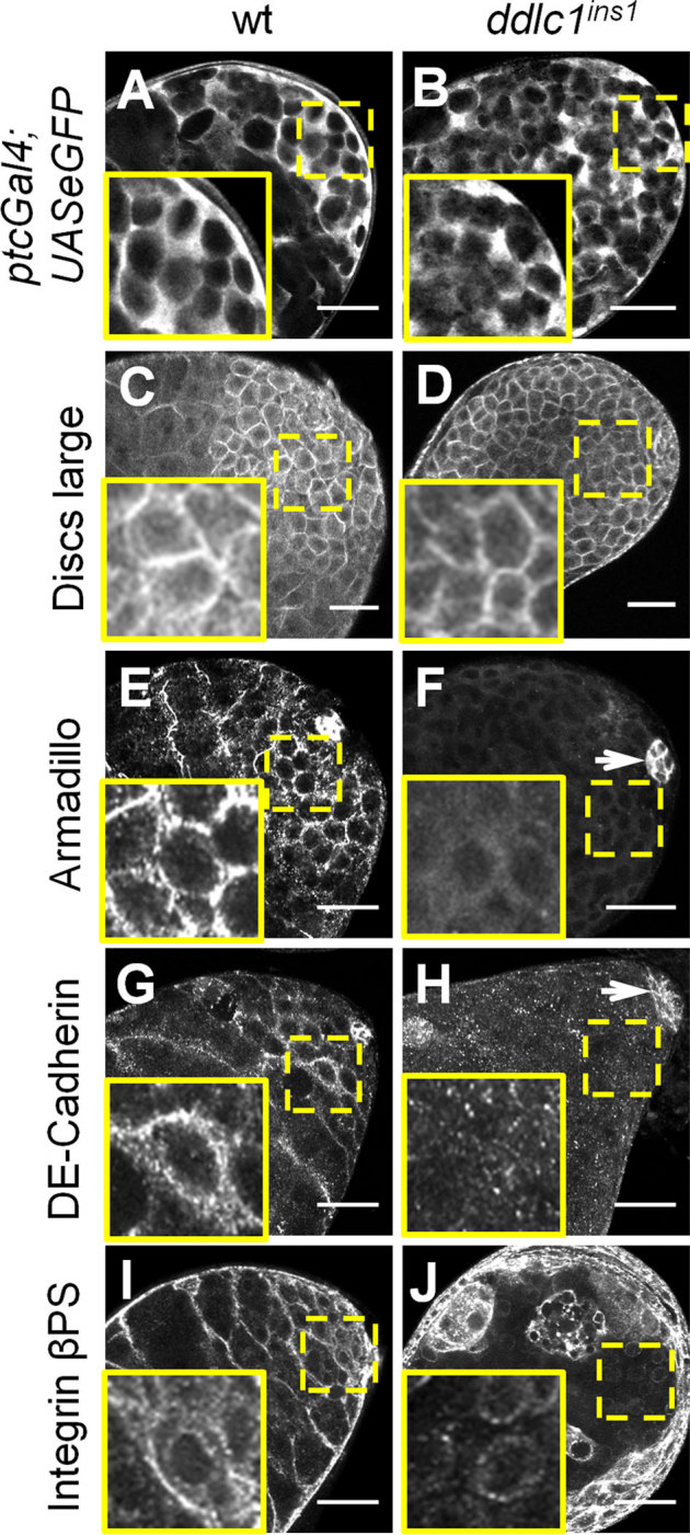
Stem cell progeny often undergo transit amplifying divisions before differentiation. In Drosophila, a spermatogonial precursor divides four times within an enclosure formed by two somatic-origin cyst cells, before differentiating into spermatocytes. Although germline and cyst cell-intrinsic factors are known to regulate these divisions, the mechanistic details are unclear. Here, we show that loss of dynein-light-chain-1 (DDLC1/LC8) in the cyst cells eliminates bag-of-marbles (bam) expression in spermatogonia, causing gonial cell hyperplasia in Drosophila testis. The phenotype is dominantly enhanced by Dhc64C (cytoplasmic Dynein) and didum (Myosin V) loss-of-function alleles. Loss of DDLC1 or Myosin V in the cyst cells also affects their differentiation. Furthermore, cyst cell-specific loss of ddlc1 disrupts Armadillo, DE-cadherin and Integrin-βPS localizations in the cyst. Together, these results suggest that Dynein and Myosin V activities, and independent DDLC1 functions in the cyst cells organize the somatic microenvironment that regulates spermatogonial proliferation and differentiation.
Figures
Similar articles
-
Cytoplasmic dynein-dynactin complex is required for spermatid growth but not axoneme assembly in Drosophila.Mol Biol Cell. 2004 May;15(5):2470-83. doi: 10.1091/mbc.e03-11-0848. Epub 2004 Mar 12. Mol Biol Cell. 2004. PMID: 15020714 Free PMC article.
-
Cytoplasmic dynein (ddlc1) mutations cause morphogenetic defects and apoptotic cell death in Drosophila melanogaster.Mol Cell Biol. 1996 May;16(5):1966-77. doi: 10.1128/MCB.16.5.1966. Mol Cell Biol. 1996. PMID: 8628263 Free PMC article.
-
Somatic ERK activation during transit amplification is essential for maintaining the synchrony of germline divisions in Drosophila testis.Open Biol. 2018 Jul;8(7):180033. doi: 10.1098/rsob.180033. Open Biol. 2018. PMID: 30045884 Free PMC article.
-
The Germline Linker Histone dBigH1 and the Translational Regulator Bam Form a Repressor Loop Essential for Male Germ Stem Cell Differentiation.Cell Rep. 2017 Dec 12;21(11):3178-3189. doi: 10.1016/j.celrep.2017.11.060. Cell Rep. 2017. PMID: 29241545
-
Autophagy is required for spermatogonial differentiation in the Drosophila testis.Biol Futur. 2022 Jun;73(2):187-204. doi: 10.1007/s42977-022-00122-7. Epub 2022 Jun 7. Biol Futur. 2022. PMID: 35672498 Review.
Cited by
-
Investigating spermatogenesis in Drosophila melanogaster.Methods. 2014 Jun 15;68(1):218-27. doi: 10.1016/j.ymeth.2014.04.020. Epub 2014 May 2. Methods. 2014. PMID: 24798812 Free PMC article. Review.
-
Somatic PI3K activity regulates transition to the spermatocyte stages in Drosophila testis.J Biosci. 2017 Jun;42(2):285-297. doi: 10.1007/s12038-017-9678-5. J Biosci. 2017. PMID: 28569252
-
Three RNA binding proteins form a complex to promote differentiation of germline stem cell lineage in Drosophila.PLoS Genet. 2014 Nov 20;10(11):e1004797. doi: 10.1371/journal.pgen.1004797. eCollection 2014 Nov. PLoS Genet. 2014. PMID: 25412508 Free PMC article.
-
A polycistronic locus encodes both a tumor-suppressive microRNA and a long non-coding RNA regulating testis morphology in Drosophila.bioRxiv [Preprint]. 2025 May 16:2024.10.10.617551. doi: 10.1101/2024.10.10.617551. bioRxiv. 2025. PMID: 39416153 Free PMC article. Preprint.
-
CG8005 Mediates Transit-Amplifying Spermatogonial Divisions via Oxidative Stress in Drosophila Testes.Oxid Med Cell Longev. 2020 Oct 27;2020:2846727. doi: 10.1155/2020/2846727. eCollection 2020. Oxid Med Cell Longev. 2020. PMID: 33193998 Free PMC article.
References
-
- Lajtha L. G. Stem cell concepts. Differentiation 14, 23–34 (1979). - PubMed
-
- Ingber D. E. Cancer as a disease of epithelial-mesenchymal interactions and extracellular matrix regulation. Differentiation 70, 547–560 (2002). - PubMed
-
- Stover D. G., Bierie B. & Moses H. L. A delicate balance: TGF-beta and the tumor microenvironment. J Cell Biochem 101, 851–861 (2007). - PubMed
-
- Fuller M. T. in The Development of Drosophila melanogaster Vol. 1 (edBate M., ed. ) 71–147 (Cold Spring Harbor Laboratory Press, Cold Spring Harbor, NY, New York, 1993).
-
- Gonczy P. & DiNardo S. The germ line regulates somatic cyst cell proliferation and fate during Drosophila spermatogenesis. Development 122, 2437–2447 (1996). - PubMed
Publication types
MeSH terms
Substances
Grants and funding
LinkOut - more resources
Full Text Sources
Molecular Biology Databases
Research Materials
Miscellaneous
