

SACK-expanded hair follicle stem cells display asymmetric nuclear Lgr5 expression with non-random sister chromatid segregation
- PMID: 22355691
- PMCID: PMC3240967
- DOI: 10.1038/srep00176
SACK-expanded hair follicle stem cells display asymmetric nuclear Lgr5 expression with non-random sister chromatid segregation
Abstract
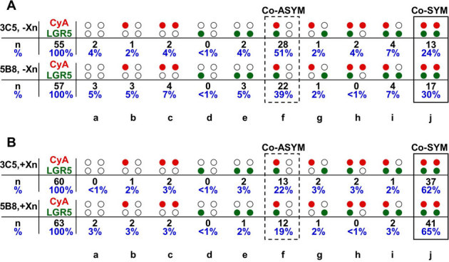
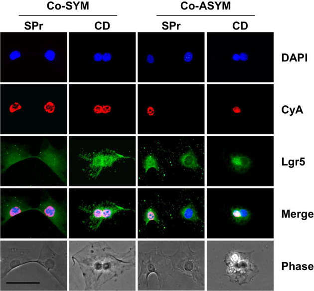
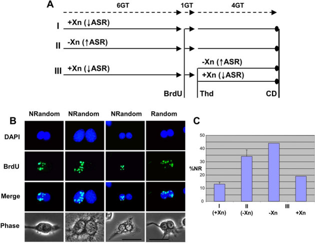
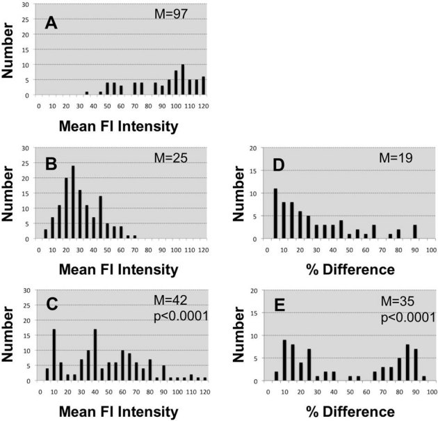
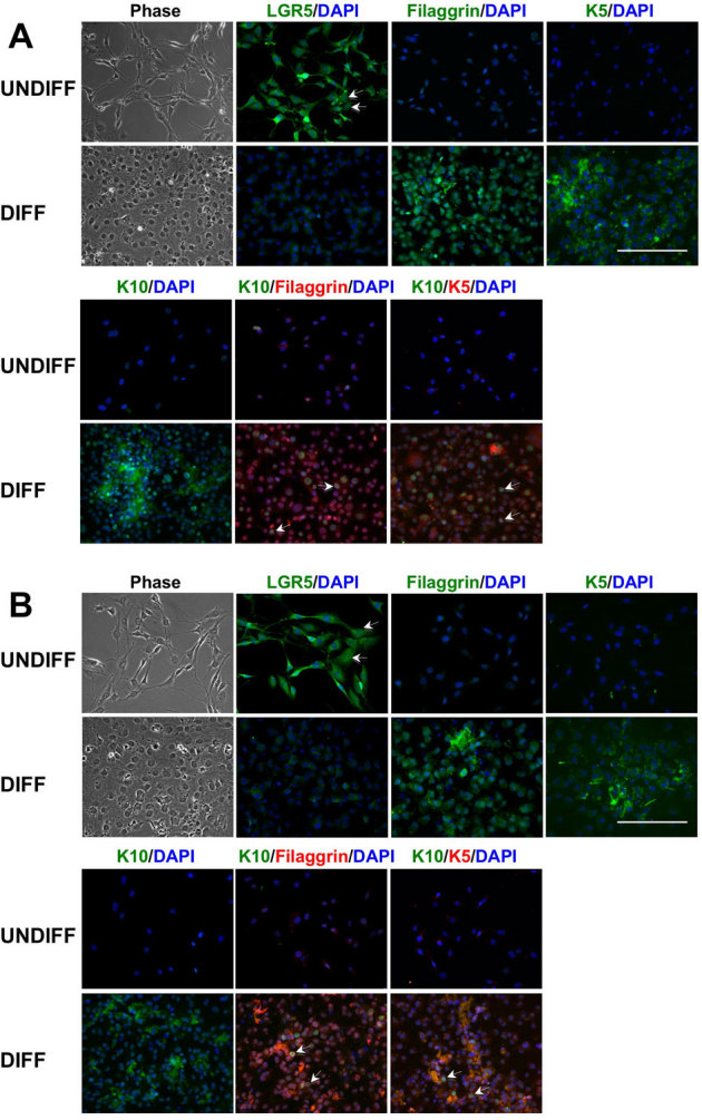
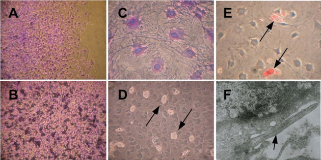
We investigated the properties of clonally-expanded mouse hair follicle stem cells (HF-SCs) in culture. The expansion method, suppression of asymmetric cell kinetics (SACK), is non-toxic and reversible, allowing evaluation of the cells' asymmetric production of differentiating progeny cells. A tight association was discovered between non-random sister chromatid segregation, a unique property of distributed stem cells (DSCs), like HF-SCs, and a recently described biomarker, Lgr5. We found that nuclear Lgr5 expression was limited to the HF-SC sister of asymmetric self-renewal divisions that retained non-randomly co-segregated chromosomes, which contain the oldest cellular DNA strands, called immortal DNA strands. This pattern-specific Lgr5 association poses a potential highly specific new biomarker for delineation of DSCs. The expanded HF-SCs also maintained the ability to make differentiated hair follicle cells spontaneously, as well as under conditions that induced cell differentiation. In future human cell studies, this capability would improve skin grafts and hair replacement therapies.
Figures
Similar articles
-
Higher 5-hydroxymethylcytosine identifies immortal DNA strand chromosomes in asymmetrically self-renewing distributed stem cells.Proc Natl Acad Sci U S A. 2013 Oct 15;110(42):16862-7. doi: 10.1073/pnas.1310323110. Epub 2013 Sep 30. Proc Natl Acad Sci U S A. 2013. PMID: 24082118 Free PMC article.
-
Decreased H3K27 and H3K4 trimethylation on mortal chromosomes in distributed stem cells.Cell Death Dis. 2014 Dec 4;5(12):e1554. doi: 10.1038/cddis.2014.522. Cell Death Dis. 2014. PMID: 25476902 Free PMC article.
-
Molecular cloaking of H2A.Z on mortal DNA chromosomes during nonrandom segregation.Stem Cells. 2011 Oct;29(10):1620-7. doi: 10.1002/stem.707. Stem Cells. 2011. PMID: 21905168
-
Centromere assembly and non-random sister chromatid segregation in stem cells.Essays Biochem. 2020 Sep 4;64(2):223-232. doi: 10.1042/EBC20190066. Essays Biochem. 2020. PMID: 32406510 Review.
-
Wnt signaling, lgr5, and stem cells in the intestine and skin.Am J Pathol. 2009 Mar;174(3):715-21. doi: 10.2353/ajpath.2009.080758. Epub 2009 Feb 5. Am J Pathol. 2009. PMID: 19197002 Free PMC article. Review.
Cited by
-
Higher 5-hydroxymethylcytosine identifies immortal DNA strand chromosomes in asymmetrically self-renewing distributed stem cells.Proc Natl Acad Sci U S A. 2013 Oct 15;110(42):16862-7. doi: 10.1073/pnas.1310323110. Epub 2013 Sep 30. Proc Natl Acad Sci U S A. 2013. PMID: 24082118 Free PMC article.
-
When stem cells grow old: phenotypes and mechanisms of stem cell aging.Development. 2016 Jan 1;143(1):3-14. doi: 10.1242/dev.130633. Development. 2016. PMID: 26732838 Free PMC article. Review.
-
Adult hair follicle stem cells do not retain the older DNA strands in vivo during normal tissue homeostasis.Chromosome Res. 2013 May;21(3):203-12. doi: 10.1007/s10577-013-9355-y. Chromosome Res. 2013. PMID: 23681654 Free PMC article. Review.
-
In vitro expansion of the mammary stem/progenitor cell population by xanthosine treatment.BMC Cell Biol. 2012 Jun 14;13:14. doi: 10.1186/1471-2121-13-14. BMC Cell Biol. 2012. PMID: 22698263 Free PMC article.
-
Decreased H3K27 and H3K4 trimethylation on mortal chromosomes in distributed stem cells.Cell Death Dis. 2014 Dec 4;5(12):e1554. doi: 10.1038/cddis.2014.522. Cell Death Dis. 2014. PMID: 25476902 Free PMC article.
References
-
- Paus R. & Cotsarelis G. The biology of hair follicles. New Eng. J. Med. 341, 491–497 (1999). - PubMed
-
- Taylor G., Lehrer M. S., Jensen P. J., Sun T.-T., and Lavker R. M. Involvement of follicular stem cells in forming not only the follicle but also the epidermis. Cell 102, 451–461 (2000). - PubMed
-
- Oshima H., Rochat A., Kedzia C., Kobayashi K., & Barrandon Y. Morphogenesis and renewal of hair follicles from adult multipotent stem cells. Cell 104, 233–245 (2001). - PubMed
-
- Celso. et al. Characterization of bipotential epidermal progenitors derived from human sebaceous gland: contrasting roles of c-myc and β-catenin. .Stem Cells 26, 1241–1252 (2008). - PubMed
Publication types
MeSH terms
Substances
Grants and funding
LinkOut - more resources
Full Text Sources
Other Literature Sources
Medical
Research Materials
Miscellaneous
