. 2011:1:180.
doi: 10.1038/srep00180. Epub 2011 Dec 5.

Potential role of LMP2 as tumor-suppressor defines new targets for uterine leiomyosarcoma therapy

Affiliations

Potential role of LMP2 as tumor-suppressor defines new targets for uterine leiomyosarcoma therapy

Takuma Hayashi et al. Sci Rep. 2011.

Abstract

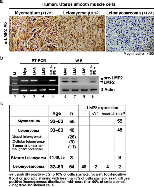
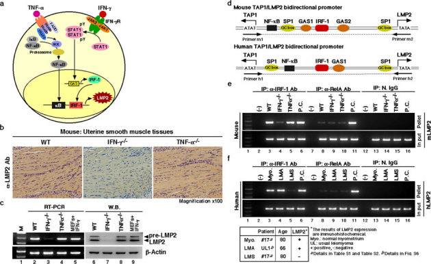
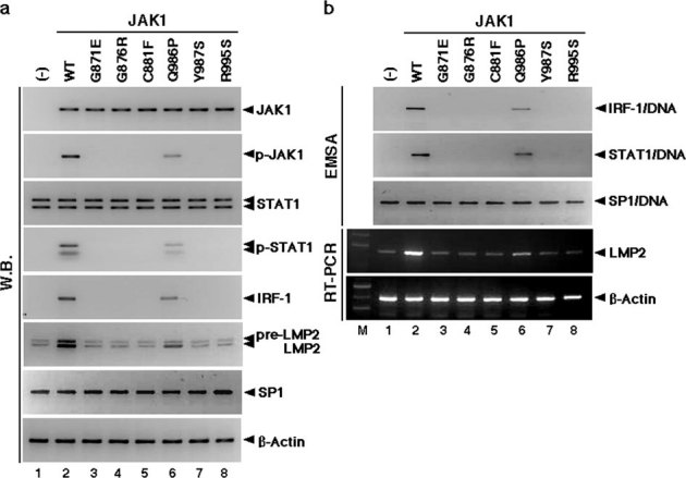
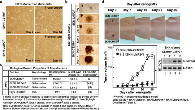
Although the majority of smooth muscle neoplasms found in the uterus are benign, uterine leiomyosarcoma (LMS) is extremely malignant, with high rates of recurrence and metastasis. We earlier reported that mice with a homozygous deficiency for LMP2, an interferon (IFN)-γ-inducible factor, spontaneously develop uterine LMS. The IFN-γ pathway is important for control of tumor growth and invasion and has been implicated in several cancers. In this study, experiments with human and mouse uterine tissues revealed a defective LMP2 expression in human uterine LMS that was traced to the IFN-γ pathway and the specific effect of JAK-1 somatic mutations on the LMP2 transcriptional activation. Furthermore, analysis of a human uterine LMS cell line clarified the biological significance of LMP2 in malignant myometrium transformation and cell cycle, thus implicating LMP2 as an anti-tumorigenic candidate. This role of LMP2 as a tumor suppressor may lead to new therapeutic targets in human uterine LMS.


Figures

Figure 1
Figure 1. Defect in LMP2 expression in human uterine leiomyosarcoma (LMS) tissue.
(a) Immunohistochemistry (IHC) of LMP2 in normal myometrium (patient #17a), uterine leiomyoma (LMA, patient UL1b) and uterine LMS (patient #17a) tissues located in the same tissue. For all samples, 5 μm sections of tissue were stained with anti-LMP2 antibody and revealed by peroxidase-conjugated anti-rabbit IgG antibody. (magnification x100) a,bDetails of patients with LMA or LMS are shown in Table. S1 and Fig. S6. (b) Cytosolic extracts were prepared from normal human myometrium (patient #17a), uterine leiomyoma (LMA, patient UL1b), and uterine LMS (patient #17a) tissues. Extracts of 50 μg were resolved by 10% sodium dodecyl sulfate-polyacrylamide gel electrophoresis (SDS-PAGE). The levels of LMP2 and β-actin were examined by immunoblot analysis with appropriate antibodies. Examinations of mRNA expression for LMP2, β2-m, and β-actin in normal myometrium (Myo.), uterine LMA, and uterine LMS were performed by reverse transcription-polymerase chain reaction (RT-PCR) with the appropriate primers indicated in the Materials and Methods section. The DNA products amplified by RT-PCR were loaded onto agarose gels. aDetails are shown in Table S1 and Table S2, bdetails are shown in Fig. S6. (c) IHC experiments individually performed at several medical facilities revealed a marked loss in the ability to induce LMP2 expression in human uterine LMS tissues compared to that in normal myometrium located in the same tissue section, as well as to that in LMA tissues. Normal total: 55 cases, LMA total: 48 cases, Bizarre Leiomyoma total: 3 cases, LMS total: 54 cases. The experiments were performed three times with similar results.
Figure 2
Figure 2. Key role of the IFN-γ-pathway in LMP2 expression in normal myometrium.
(a) Key role of the signaling pathway on LMP2 expression. (b) Immunohistochemical experiments with myometrium tissue sections derived from wild-type, IFN-γ-deficient, and TNF-α-deficient mice (2 months old) were carried out. (magnification x100) The results revealed that the IFN-γ signaling cascade was required for basal LMP2 expression. (c) Western blotting and RT-PCR experiments with myometrium tissue sections derived from wild-type and IFN-γ- and TNF-α-deficient mice (2 months old) were also performed. The results showed that IFN-γ-deficient mice had markedly reduced LMP2 levels in myometrium tissues. These findings support the notion that the IFN-γ pathway plays a key role in basal LMP2 expression. (d) Schematic representation of the LMP2/TAP1 bidirectional promoter, including NF-κB and IRF-1 binding sites. Chromatin immunoprecipitation (ChIP) analysis with antibodies against RelA (NF-κBp65) and IRF-1 was carried out. (e) ChIP assays showing that although mouse genomic DNA of the Lmp2 enhancer/promoter region was markedly amplified using immunoprecipitated TNF-α-deficient myometrium tissue with anti-IRF-1 antibody, amplified products were not detected using immunoprecipitated IFN-γ-deficient myometrium tissue with anti-IRF-1 antibody. The mouse genomic DNA of the LMP2 enhancer/promoter region was unclearly amplified using the immunoprecipitated materials with anti-RelA antibody. Positive control (P.C.): IFN-γ-treated mouse embryonic fibroblasts (lane 6), TNF-α-treated mouse splenocytes (lane 11). (f) ChIP assays showing that although human genomic DNA of the LMP2 enhancer/promoter region was markedly amplified using immunoprecipitated LMA tissue as well as normal myometrium tissue with anti-IRF-1 antibody, no-DNA amplification was detected in the immunoprecipitated LMS sample with anti-IRF-1 antibody. The DNA of the LMP2 enhancer/promoter region was not clearly amplified using any immunoprecipitated myometrium tissue materials with anti-RelA antibody. Positive control (P.C.): IFN-γ-treated mouse embryonic fibroblasts (lane 6), TNF-α-treated mouse splenocytes (lane 11). N.IgG, normal rabbit anti-serum was used as a negative control antibody. The experiments were performed four times with similar results.
Figure 3
Figure 3. Somatic JAK1 mutations prevent LMP2 expression.
(a) JAK1 mutants resulted in defective activation of downstream IFN-γ pathways, as described in Table S3. Activation of JAK1, STAT1, and IRF-1 for LMP2 expression by IFN-γ in cells expressing JAK1WT and JAK1 mutants. Protein lysates of JAK1-deficient cells expressing wild-type and JAK1 mutants were analyzed by Western blotting (W.B.) using appropriate antibodies. JAK1 and STAT1 phosphorylation after 15 minutes of stimulation was consistently increased in cells expressing JAK1WT and JAK1G986P, but phosphorylated proteins were not detected in cells expressing other JAK1 mutants. IRF-1 and LMP2 expression was markedly activated in cells expressing JAK1WT and JAK1G986P. There was no difference in the expression of JAK1, STAT1, SP1, or β-Actin among cells expressing wild-type and JAK1 mutants. (b) Defective DNA-binding activities of IRF-1 and STAT1 suggest that LMP2 expression is attributable to mutations in the catalytic domains of JAK1. DNA-binding activities of SP1 were detected in all tested samples. RT-PCR supported W.B. results. The experiments were performed three times with similar results. Direct sequencing demonstrated that the mutations detected in the catalytic sites were indeed somatic mutations (details in Supplementary Fig. S7 online).
Figure 4
Figure 4. Biological activity of hLMP2 in uterine leiomyosarcoma (LMS).
(a) Phase-contrast micrographs of the parental transformed SKN-CEM9 (T type) clone and flat revertants of the SKN-LMP2 (F type) clone. (magnification x100) (b) Phase-contrast micrographs of the colony formations of the SKN-CEM9 (T type) clone and SKN-LMP2 (F type) clone in soft agar. (magnification x40) (c) The table indicates the biological properties of the transfectants, whose details are described in Table 1 and Supplementary Table 5. (d) Changes in the human uterine LMS cell line, SKN transfectants, the SKN-CEM9 (T type) clone, and the SKN-LMP2 (F type) clone xenograft volumes in mice (n = 8). Representative photographs of xenografts in mice (Upper panel). Tumor growth of SKN-LMP2 was markedly reduced in comparison with that of the control transfectant SKN-CEM9 (T type) clone. Tumor growth kinetics after subcutaneous injection of the SKN-CEM9 (T type) clone and SKN-LMP2 (F type) clone (Lower panel). RT-PCR experiments reveal hLMP2 mRNA expression in tumors (Lower right panel). The experiments were performed four times with similar results.

Similar articles

Cited by

References

    1. Zaloudek C. & Hendrickson M. R. Mesenchymal tumors of the uterus, in Kurman RJ. (ed): Blaustein's Pathology of the Female Genital Tract (ed 5). New York, Springer-Verlag 561–578 (2002).
    1. Gadducci A., et al. Uterine leiomyosarcoma: analysis of treatment failures and survival. Gynecol. Oncol. 62, 25–32 (1996). - PubMed
    1. Nordal R. & Thoresen S. Uterine sarcomas in Norway 1956–1992: incidence, survival and mortality. Eur. J. Cancer 33, 907–311 (1997). - PubMed
    1. Brooks S. E., Zhan M., Cote T. & Baquet C. R. Surveillance, epidemiology, and end results analysis of 2677 cases of uterine sarcoma 1989–1999. Gynecol. Oncol. 93, 204–208 (2004). - PubMed
    1. Dusenbery K. E., Potish R. A., Argenta P. A. & Judson P. L. On the apparent failure of adjuvant pelvic radiotherapy to improve survival for women with uterine sarcomas confined to the uterus. Am. J. Clin. Oncol. 28, 295–300 (2005). - PubMed

Publication types