. 2011:1:203.
doi: 10.1038/srep00203. Epub 2011 Dec 21.

The regulatory role of long-term depression in juvenile and adult mouse ocular dominance plasticity

Affiliations

The regulatory role of long-term depression in juvenile and adult mouse ocular dominance plasticity

Kaiyun Yang et al. Sci Rep. 2011.

Erratum in

  • Sci Rep. 2013;3:1442. Taghibiglou, Changiz [added]

Abstract

The study of experience-dependent ocular dominance (OD) plasticity has greatly contributed to the understanding of visual development. During the critical period, preventing input from one eye results in a significant impairment of vision, and loss of cortical responsivity via the deprived eye. Residual ocular dominance plasticity has recently been observed in adulthood. Accumulating evidence suggests that OD plasticity involves N-methyl-(D)-aspartate receptor (NMDAR)-dependent long-term depression (LTD). Here we report that the administration of a selective LTD antagonist prevented the ocular dominance shift during the critical period. The NMDAR co-agonist D-serine facilitated adult visual cortical LTD and the OD shift in short-term monocularly deprived (MD) adult mice. When combined with reverse suture, D-serine proved effective in restoring a contralaterally-dominated visual input pattern in long-term MD mice. This work suggests LTD as a key mechanism in both juvenile and adult ocular dominance plasticity, and D-serine as a potential therapeutic in human amblyopic subjects.


Figures
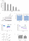
Figure 1
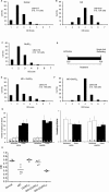
Figure 1. Correlation between LTD magnitude and the critical period; LTD blockade.
(A) Local field potential recording showed that the magnitude of LFS-induced LTD declined with development, and was much diminished in adulthood. (B) Pretreatment of the slices with Tat-GluR23Y (1 μM; 30 min), but not scrambled control (Tat-sGluR23Y, 1 μM; 30 min), prevented the NMDA-mediated reduction in AMPARs in response to bath application of NMDA (20 μM; 5 min) in visual cortical slices (around P25), without altering the basal level of surface AMPARs, or GABAA receptors. GSK3b was probed as a control for specific biotinylation of surface proteins. (C) Quantification of surface AMPARs and GABAA receptors after chemical LTD induction. A marked reduction of surface GluR2 subunits was observed both in the absence of Tat-GluR23Y (81.96 ± 4.42 % of control, n = 3; p<0.05) and with pretreatment with scrambled Tat-GluR23Y (79.06 ± 4.13 % of control, n = 3; p<0.05), but not with pretreatment with Tat-GluR23Y (94.39 ± 6.92 % of control, n = 3; p = 0.45). The expression of GABAA β2/3 on the cell surface was not changed by NMDA treatment (96.19 ± 4.81 %, n = 3; p = 0.46), neither Tat-GluR23Y (108.10 ± 13.00 %, n = 3; p = 0.56) nor scrambled Tat-GluR23Y (95.91 ± 5.26 %, n = 3; p = 0.47) pretreatment altered that effect. Asterisk, p < 0.05. Error bars reflect SEM. (D) Bath application of Tat-GluR23Y blocked NMDAR-dependant LTD of field recordings in primary visual cortical slices (around P25). Red diamond, GluR23A (n = 6); green circle, GluR23Y (n = 7); blue circle, control LTD (n = 7); light blue triangle, D-APV treatment (n = 5). (D) Inclusion of GluR23Y (100 μM; n = 7), but not GluR23A (100 μM; n = 7) in patch electrodes blocked LTD of excitatory postsynaptic currents (EPSCs) in whole-cell recordings. (F) Representative fEPSPs and EPSCs taken just before the LFS (1) and near the end of recording (2) as indicated in both Fig 1D and 1E. (G) Summary of results obtained under field recording and whole-cell recording conditions. Each bar represents the average of normalized fEPSPs or EPSCs recorded in the last 5 min under conditions as labeled on the X-axis. Asterisk, p < 0.05. Error bar represents SEM.
Figure 2
Figure 2. GluR23Y peptide administration prevents the ocular dominance shift in vivo.
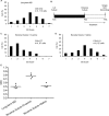
(A) The OD distribution favored the contralateral eye in normal non-deprived (ND) mice. (B) Monocular deprivation shifted the distribution towards the ipsilateral eye. (C) Tat-GluR23Y peptide had no influence on OD in ND mice. (D) Tat-GluR23Y or Tat-GluR23A (10 nmol/g; i.p.) was administered daily during the four-day MD period. (E) Tat-GluR23Y (10 nmol/g; i.p.) prevented the OD shift following MD (p = 0.26 compared to nondeprived mice). (F) Tat-GluR23A did not prevent the OD shift following MD. (G) Tat-GluR23Y had no acute influence on the spontaneous activity and evoked response in the binocular zone of the primary visual cortex. The number of spikes was counted with or without the presence of the visual stimulus (a moving grating), 1 h or 2 h after i.p. injection of saline or peptide. The Y-axis shows the number of spikes obtained. White bars represent spontaneous activity and blue bars represent evoked activity. Bars from left to right represent before peptide/saline administration; 1 h after administration and 2 h after administration. For statistical analysis, spontaneous and evoked spikes at 1 h and 2 h are normalized against the spontaneous or evoked activity at 0 h at the same site on the same animal. The same normalization was done for animals treated with peptide or saline. (H) Data summary of single-unit recordings in juvenile mice. The dots represent individual animals. The horizontal bar represents the mean of the CBI for each group.
Figure 3
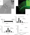
Figure 3. Local infusion of Tat-GluR23Y in the primary visual cortex blocks the OD shift.
(A) FITC-conjugated Tat-GluR23Y (2 µM) was delivered using osmotic minipumps. After 24 h, the fluorescent dye was detected throughout the visual cortex, but not in the brain stem or on the contralateral side of the brain. The left panel was taken under transmitted light, the right panel was taken under fluorescence. Scale bar (1 mm) is illustrated at the bottom right. The binocular zone is highlighted. (B) Experimental schedule for intracortical infusion of the peptides. (C) Local infusion of Tat-GluR23Y prevented the OD shift (0.76 ± 0.01, n = 5; p = 0.82 compared to ND mice and p = 0.004 compared to MD mice). (D) Local infusion of Tat-GluR23A did not prevent the OD shift (CBI = 0.46 ± 0.01, n = 5; p = 0.005 compared to ND mice, and p = 0.33 compared to MD mice). (E) data Summary of the binocularity index results under the various conditions studied.
Figure 4
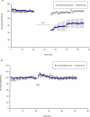
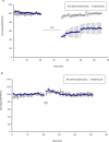
Figure 4. D-serine selectively facilitates the induction of LTD in adult visual cortical slices.
(A) D-serine facilitated LTD in adult visual cortical slices. 20 minutes of D-serine (20 µM) incubation facilitated the induction of LTD (66±12% of baseline, n = 4; p< 0.01) after delivering low-frequency stimulation (1 Hz, 900 stimuli). Error bars reflect SEM. (B) D-serine did not facilitate LTP in adult visual cortical slices. 20 minutes of D-serine incubation (concentrations from 20µM up to 200 µM) did not affect the induction of LTP (98±4%, n = 5) after delivering theta-burst stimulation.
Figure 5
Figure 5. D-serine enhances ocular dominance plasticity in adult mice.
(A) Normal adult mice demonstrated a contralateral biased OD distribution (CBI = 0.78±0.01, n = 5). (B) single-unit recordings revealed no ocular dominance shift in monocularly deprived adult mice (CBI = 0.78±0.01, n = 5, p = 0.64 compared to normal adult mice). (C) D-serine itself (600 mg/kg, bid, s.c.) did not induce an ocular dominance shift compared to untreated adult mice (CBI = 0.74±0.01, n = 5, p = 0.03). (D) Experimental schedule for single-unit recording experiments. (E) D-serine (600 mg/kg, bid, s.c.) reinstated the ocular dominance shift after monocular deprivation in adult mice as is shown in the ocular dominance score distribution (CBI = 0.56±0.05, n = 5, p<0.01 compared to normal mice). (F) The saline treatment group did not show a corresponding shift in OD distribution (CBI = 0.77±0.01, n = 5, p = 0.23 compared to normal mice). (G) GluR23Y (10 nmol/g, i.p.) prevented the effect of D-serine (600 mg/kg, bid, s.c.) in monocularly deprived adult mice (CBI = 0.68, n = 4). (H) Data summary of the binocularity index results in the various conditions studied in adult mice.
Figure 6
Figure 6. D-serine enhances visual cortical plasticity in adult mice.
(A) Experimental schedule for single-unit recording experiments in adult mice that received local infusion treatment. (B) D-serine-infused (50 μM) mice showed a significant ocular dominance shift (CBI = 0.47±0.01, n = 3, p<0.01 compared to normal mice). (C) Saline-infused mice demonstrated an OD distribution favoring the contralateral eye (CBI = 0.78±0.01, n = 3). (D) Data summary of the binocularity index in the D-serine intracortical infusion study.
Figure 7
Figure 7. D-serine promotes the restoration of normal binocularity in reverse sutured mice.
(A) Long-term monocular deprivation results in an ocular dominance shift favoring the open eye into adulthood (CBI = 0.50±0.05, n = 5). (B) Experimental schedule for single-unit recording experiments in reverse sutured animals. (C) D-serine (600 mg/kg, bid, s.c.) promoted recovery of visual input from a previously deprived eye in reverse sutured mice (CBI = 0.77±0.03, n = 4). (D) Saline treated RS mice still showed dominance by the previously open eye (CBI = 0.51±0.03, n = 4). (E) CBI summary in long-term deprived mice, and in RS mice treated with D-serine or saline.

Similar articles

Cited by

References

    1. Hofer S. B., Mrsic-Flogel T. D., Bonhoeffer T. & Hubener M. Lifelong learning: ocular dominance plasticity in mouse visual cortex. Current opinion in neurobiology 16, 451–459 (2006). - PubMed
    1. Karmarkar U. R. & Dan Y. Experience-dependent plasticity in adult visual cortex. Neuron 52, 577–585 (2006). - PubMed
    1. Prusky G. T., Silver B. D., Tschetter W. W., Alam N. M. & Douglas R. M. Experience-dependent plasticity from eye opening enables lasting, visual cortex-dependent enhancement of motion vision. J Neurosci 28, 9817–9827 (2008). - PMC - PubMed
    1. Hensch T. K. et al. Local GABA circuit control of experience-dependent plasticity in developing visual cortex. Science (New York, N.Y 282, 1504–1508 (1998). - PMC - PubMed
    1. Cynader M. & Mitchell D. E. Prolonged sensitivity to monocular deprivation in dark-reared cats. Journal of neurophysiology 43, 1026–1040 (1980). - PubMed

Publication types