. 2012 Apr 2;9(4):946-56.
doi: 10.1021/mp200553x. Epub 2012 Mar 20.

Dendritic cell targeted chitosan nanoparticles for nasal DNA immunization against SARS CoV nucleocapsid protein

Affiliations

Dendritic cell targeted chitosan nanoparticles for nasal DNA immunization against SARS CoV nucleocapsid protein

Dharmendra Raghuwanshi et al. Mol Pharm. .

Abstract

This work investigates the formulation and in vivo efficacy of dendritic cell (DC) targeted plasmid DNA loaded biotinylated chitosan nanoparticles for nasal immunization against nucleocapsid (N) protein of severe acute respiratory syndrome coronavirus (SARS-CoV) as antigen. The induction of antigen-specific mucosal and systemic immune response at the site of virus entry is a major challenge for vaccine design. Here, we designed a strategy for noninvasive receptor mediated gene delivery to nasal resident DCs. The pDNA loaded biotinylated chitosan nanoparticles were prepared using a complex coacervation process and characterized for size, shape, surface charge, plasmid DNA loading and protection against nuclease digestion. The pDNA loaded biotinylated chitosan nanoparticles were targeted with bifunctional fusion protein (bfFp) vector for achieving DC selective targeting. The bfFp is a recombinant fusion protein consisting of truncated core-streptavidin fused with anti-DEC-205 single chain antibody (scFv). The core-streptavidin arm of fusion protein binds with biotinylated nanoparticles, while anti-DEC-205 scFv imparts targeting specificity to DC DEC-205 receptor. We demonstrate that intranasal administration of bfFp targeted formulations along with anti-CD40 DC maturation stimuli enhanced magnitude of mucosal IgA as well as systemic IgG against N protein. The strategy led to the detection of augmented levels of N protein specific systemic IgG and nasal IgA antibodies. However, following intranasal delivery of naked pDNA no mucosal and systemic immune responses were detected. A parallel comparison of targeted formulations using intramuscular and intranasal routes showed that the intramuscular route is superior for induction of systemic IgG responses compared with the intranasal route. Our results suggest that targeted pDNA delivery through a noninvasive intranasal route can be a strategy for designing low-dose vaccines.


Figures

Figure 1
Figure 1
Schematic representation of dendritic cell targeting strategy using bifunctional fusion protein (bfFp). The bfFp is a recombinant fusion protein consisting of truncated core-streptavidin fused with anti-DEC-205 single chain antibody (scFv). The core-streptavidin arm of fusion protein binds with biotinylated nanoparticles while anti-DEC-205 scFv imparts targeting specificity to DC DEC-205 receptor.
Figure 2
Figure 2
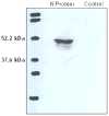
Analysis of recombinant N protein expression. The COS-1 cells were transiently transfected with pVAXN or pVAX-1 (control). N protein expression on cell lysate was analyzed by probing Western blot with mouse anti-N-specific monoclonal (19C7).
Figure 3
Figure 3
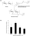
(a) Schematic for the synthesis of biotinylated chitosan. (b) Estimation of chitosan modification using fluorescamine assay. The extent of biotinylation of the amine groups of chitosan was determined using fluorescamine, which reacts with primary amine groups. The decrease in fluorescence intensity is proportional to the percentage of biotinylated amine groups.
Figure 4
Figure 4
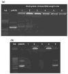
Formulation of pVAXN loaded biotinylated chitosan nanoparticles. (a) Agarose gel analysis of pVAXN DNA loaded biotinylated chitosan nanoparticles prepared at different weight ratios of B-chitosan to pVAXN DNA (1 to 6). (b) Agarose gel electrophoresis of DNAse I digested naked pVAXN and nanoencapsulated pVAXN. Lane 1: pVAXN (30 min); Lane 2: nanoencapsulated pVAXN (30 min); Lane 3: pVAXN (60 min); Lane 4: nanoencapsulated pVAXN (60 min); Lane 5: Lane 4 digested with chitosanase and lysozyme.
Figure 5
Figure 5
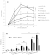
(a) Analysis of time-dependent serum IgG antibody against SARS-CoV N protein following IN and IM immunization with different vaccine formulations. Balb/c mice (5 mice per group) were immunized with 5 μg of pVAXN on day 1 and day 21. The mouse serum was collected at day 0, 14, 28, 42 and 56 post immunization and IgG against N protein was detected using ELISA. The data are presented as group mean ± SD at various time-points. ELISA was performed at 1:100 diluted sera. (b) Detection of N protein specific serum IgG isotypes in mice vaccinated with different formulations. IgG isotypes were detected in serum samples obtained on day 56. The ELISA was performed at 1:100 diluted sera. Data are presented as group mean ± SD. Statistical differences between groups are denoted as *p<0.05 and ns = no significant difference (p>0.05). The horizontal bars compare statistical difference in IgG1 between different groups.
Figure 6
Figure 6
SARS-CoV N protein specific IgA levels in the nasal washes. The nasal washes were collected on day 56 following euthanasia and assayed for the presence of N protein specific IgA using ELISA. Each sample was analyzed in triplicate and data are representative of group mean ± SD of five mice. Statistical differences between groups are denoted as *p<0.05 and ns = no significant difference (p>0.05). The horizontal bars with * denote statistical difference in IgA between different groups compared with NP+bfFp+αCD40 group (IN).
Figure 7
Figure 7
Analysis of IFN-γ levels in mice immunized with different vaccine formulations. Splenocytes obtained from a group of immunized mice (5 mice per group) were pooled and cultured in the presence of recombinant N protein (4 μg) in 96-well flat bottom plate with a final volume of 200 uL. Culture supernatants harvested after 72 hr were analyzed for IFN-γ levels using ELISA kit. Data represents mean ± SD of triplicate cultures stimulated with N protein. Statistical differences between groups are denoted as *p<0.05. * denote statistical difference in IFN-γ between different groups. IFN-γ levels obtained following stimulation with Con A were 1795–2526 pg/ml in different groups, whereas, no detectable levels were observed for splenocytes culture stimulated with media alone.

Similar articles

Cited by

References

    1. Steinman RM. Dendritic Cells In Vivo: A Key Target for a New Vaccine Science. Immunity. 2008;29(3):319–324. - PubMed
    1. Tacken PJ, De Vries IJM, Torensma R, Figdor CG. Dendritic-cell immunotherapy: From ex vivo loading to in vivo targeting. Nat Rev Immunol. 2007;7(10):790–802. - PubMed
    1. Keler T, He L, Ramakrishna V, Champion B. Antibody-targeted vaccines. Oncogene. 2007;26(25):3758–3767. - PubMed
    1. Proudfoot O, Apostolopoulos V, Pietersz GA. Receptor-mediated delivery of antigens to dendritic cells: Anticancer applications. Mol Pharm. 2007;4(1):58–72. - PubMed
    1. Inaba I, Swiggard WJ, Inaba M, Meltzer J, Mirza A, Sasagawa T, Nussenzweig MC, Steinman RM. Tissue distribution of the DEC-205 protein that is detected by the monoclonal antibody NLDC-145 I. Expression on dendritic cells and other subsets of mouse leukocytes. Cell Immunol. 1995;163(1):148–156. - PubMed

Publication types