

Discrimination of basal cell carcinoma from benign lesions based on extraction of ulcer features in polarized-light dermoscopy images
- PMID: 22356565
- PMCID: PMC3640437
- DOI: 10.1111/j.1600-0846.2011.00595.x
Discrimination of basal cell carcinoma from benign lesions based on extraction of ulcer features in polarized-light dermoscopy images
Abstract
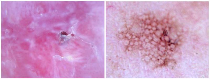
Background: Ulcers are frequently visible in magnified, cross-polarized, dermoscopy images of basal cell carcinoma. An ulcer without a history of trauma, a so-called 'atraumatic' ulcer, is an important sign of basal cell carcinoma, the most common skin cancer. Distinguishing such ulcers from similar features found in benign lesions is challenging. In this research, color and texture features of ulcers are analyzed to discriminate basal cell carcinoma from benign lesions.
Methods: Ulcers in polarized-light dermoscopy images of 49 biopsy-proven basal cell carcinomas were identified and manually selected. For 153 polarized-light dermoscopy images of benign lesions, those areas that most closely mimicked ulcers were similarly selected. Fifteen measures were analyzed over the areas of ulcers and ulcer mimics. Six of those measures were texture measures: energy, variance, smoothness, skewness, uniformity, and entropy. Nine of those measures were color measures: relative measures of red, green, and blue; chromaticity of red, green, and blue; and the ratios blue-to-green, blue-to-red, and green-to-red.
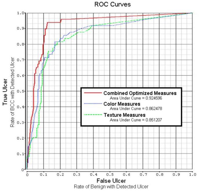
Results: A back-propagation artificial neural network was able to discriminate most of the basal cell carcinoma from benign lesions, with an area under the ROC curve as high as 92.46%, using color and texture features of ulcer areas.
Conclusion: Separation of basal cell carcinoma from benign cutaneous lesions using image analysis techniques applied to ulcers is feasible. As ulcers are a critical feature in malignant lesions, this finding may have application in the automatic detection of basal cell carcinoma.
© 2012 John Wiley & Sons A/S.
Figures

References
-
- Stoecker WV, Stolz W. Dermoscopy and the diagnostic challenge of amelanotic and hypomelanotic melanoma. Arch Dermatol. 2008;144(9):1207–10. - PubMed
-
- McFall K. Photography of dermatological conditions using polarized light. J Audiov Media Med. 1996;19(1):5–9. - PubMed
-
- Benvenuto-Andrade C, Dusza SW, Agero AL, Scope A, Rajadhyaksha M, Halpern AC, Marghoob AA. Differences between polarized light dermoscopy and immersion contact dermoscopy for the evaluation of skin lesions. Arch Dermatol. 2007;143(3):329–38. - PubMed
-
- Umbaugh SE, Moss RH, Stoecker WV. Automatic color segmentation of images with application to detection of variegated coloring in skin tumors. IEEE Eng Med Biol. 1989;8(4):43–52. - PubMed
Publication types
MeSH terms
Grants and funding
LinkOut - more resources
Full Text Sources
Medical
