Cytoplasmic BRMS1 expression in malignant melanoma is associated with increased disease-free survival
- PMID: 22356677
- PMCID: PMC3341185
- DOI: 10.1186/1471-2407-12-73
Cytoplasmic BRMS1 expression in malignant melanoma is associated with increased disease-free survival
Abstract
Background/aims: Breast cancer metastasis suppressor 1 (BRMS1) blocks metastasis in melanoma xenografts; however, its usefulness as a biomarker in human melanomas has not been widely studied. The goal was to measure BRMS1 expression in benign nevi, primary and metastatic melanomas and evaluate its impact on disease progression and prognosis.
Methods: Paraffin-embedded tissue from 155 primary melanomas, 69 metastases and 15 nevi was examined for BRMS1 expression using immunohistochemistry. siRNA mediated BRMS1 down-regulation was used to study impact on invasion and migration in melanoma cell lines.
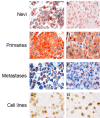
Results: A significantly higher percentage of nevi (87%), compared to primary melanomas (20%) and metastases (48%), expressed BRMS1 in the nucelus (p < 0.0001). Strong nuclear staining intensity was observed in 67% of nevi, and in 9% and 24% of the primary and metastatic melanomas, respectively (p < 0.0001). Comparable cytoplasmic expression was observed (nevi; 87%, primaries; 86%, metastases; 72%). However, a decline in cytoplasmic staining intensity was observed in metastases compared to nevi and primary tumors (26%, 47%, and 58%, respectively, p < 0.0001). Score index (percentage immunopositive celles multiplied with staining intensity) revealed that high cytoplasmic score index (≥ 4) was associated with thinner tumors (p = 0.04), lack of ulceration (p = 0.02) and increased disease-free survival (p = 0.036). When intensity and percentage BRMS1 positive cells were analyzed separately, intensity remained associated with tumor thickness (p = 0.024) and ulceration (p = 0.004) but was inversely associated with expression of proliferation markers (cyclin D3 (p = 0.008), cyclin A (p = 0.007), and p21Waf1/Cip1 (p = 0.009)). Cytoplasmic score index was inversely associated with nuclear p-Akt (p = 0.013) and positively associated with cytoplasmic p-ERK1/2 expression (p = 0.033). Nuclear BRMS1 expression in ≥ 10% of primary melanoma cells was associated with thicker tumors (p = 0.016) and decreased relapse-free period (p = 0.043). Nuclear BRMS1 was associated with expression of fatty acid binding protein 7 (FABP7; p = 0.011), a marker of invasion in melanomas. In line with this, repression of BRMS1 expression reduced the ability of melanoma cells to migrate and invade in vitro.
Conclusion: Our data suggest that BRMS1 is localized in cytoplasm and nucleus of melanocytic cells and that cellular localization determines its in vivo effect. We hypothesize that cytoplasmic BRMS1 restricts melanoma progression while nuclear BRMS1 possibly promotes melanoma cell invasion.Please see related article: http://www.biomedcentral.com/1741-7015/10/19.
Figures


Similar articles
-
The fatty acid binding protein 7 (FABP7) is involved in proliferation and invasion of melanoma cells.BMC Cancer. 2008 Sep 30;8:276. doi: 10.1186/1471-2407-8-276. BMC Cancer. 2008. PMID: 18826602 Free PMC article.
-
Importance of P-cadherin, beta-catenin, and Wnt5a/frizzled for progression of melanocytic tumors and prognosis in cutaneous melanoma.Clin Cancer Res. 2005 Dec 15;11(24 Pt 1):8606-14. doi: 10.1158/1078-0432.CCR-05-0011. Clin Cancer Res. 2005. PMID: 16361544
-
Prognostic significance of BRMS1 expression in human melanoma and its role in tumor angiogenesis.Oncogene. 2011 Feb 24;30(8):896-906. doi: 10.1038/onc.2010.470. Epub 2010 Oct 11. Oncogene. 2011. PMID: 20935672 Free PMC article.
-
Location, location, location: the BRMS1 protein and melanoma progression.BMC Med. 2012 Feb 22;10:19. doi: 10.1186/1741-7015-10-19. BMC Med. 2012. PMID: 22356729 Free PMC article. Review.
-
Selective expression of PNA-binding glycoconjugates by invasive human melanomas: a new marker of metastatic potential.Pigment Cell Res. 1994 Dec;7(6):461-4. doi: 10.1111/j.1600-0749.1994.tb00076.x. Pigment Cell Res. 1994. PMID: 7761355 Review.
Cited by
-
A germline SNP in BRMS1 predisposes patients with lung adenocarcinoma to metastasis and can be ameliorated by targeting c-fos.Sci Transl Med. 2022 Oct 5;14(665):eabo1050. doi: 10.1126/scitranslmed.abo1050. Epub 2022 Oct 5. Sci Transl Med. 2022. PMID: 36197962 Free PMC article.
-
The isolated C-terminal nuclear localization sequence of the breast cancer metastasis suppressor 1 is disordered.Arch Biochem Biophys. 2019 Mar 30;664:95-101. doi: 10.1016/j.abb.2019.01.035. Epub 2019 Jan 30. Arch Biochem Biophys. 2019. PMID: 30707944 Free PMC article.
-
MicroRNA-125a-5p inhibits invasion and metastasis of gastric cancer cells by targeting BRMS1 expression.Oncol Lett. 2018 Apr;15(4):5119-5130. doi: 10.3892/ol.2018.7983. Epub 2018 Feb 7. Oncol Lett. 2018. PMID: 29552146 Free PMC article.
-
Expression of the metastasis suppressor BRMS1 in uveal melanoma.Ecancermedicalscience. 2014 Mar 11;8:410. doi: 10.3332/ecancer.2014.410. eCollection 2014. Ecancermedicalscience. 2014. PMID: 24688598 Free PMC article.
-
Metastasis suppressors in breast cancers: mechanistic insights and clinical potential.J Mol Med (Berl). 2014 Jan;92(1):13-30. doi: 10.1007/s00109-013-1109-y. Epub 2013 Dec 6. J Mol Med (Berl). 2014. PMID: 24311119 Free PMC article. Review.
References
-
- Robertson G, Coleman A, Lugo TG. A malignant melanoma tumor suppressor on human chromosome 11. Cancer Res. 1996;56:4487–4492. - PubMed
-
- Frolova N, Edmonds MD, Bodenstine TM, Seitz R, Johnson MR, Feng R. et al.A shift from nuclear to cytoplasmic breast cancer metastasis suppressor 1 expression is associated with highly proliferative estrogen receptor-negative breast cancers. Tumour Biol. 2009;30:148–159. doi: 10.1159/000228908. - DOI - PMC - PubMed
Publication types
MeSH terms
Substances
Grants and funding
LinkOut - more resources
Full Text Sources
Medical
Miscellaneous

