Review
. 2012 Feb 22;3(1):7.
doi: 10.1186/scrt98.

Stem cell- and growth factor-based regenerative therapies for avascular necrosis of the femoral head

Affiliations
Review

Stem cell- and growth factor-based regenerative therapies for avascular necrosis of the femoral head

Lars Rackwitz et al. Stem Cell Res Ther. .

Abstract

Avascular necrosis (AVN) of the femoral head is a debilitating disease of multifactorial genesis, predominately affects young patients, and often leads to the development of secondary osteoarthritis. The evolving field of regenerative medicine offers promising treatment strategies using cells, biomaterial scaffolds, and bioactive factors, which might improve clinical outcome. Early stages of AVN with preserved structural integrity of the subchondral plate are accessible to retrograde surgical procedures, such as core decompression to reduce the intraosseous pressure and to induce bone remodeling. The additive application of concentrated bone marrow aspirates, ex vivo expanded mesenchymal stem cells, and osteogenic or angiogenic growth factors (or both) holds great potential to improve bone regeneration. In contrast, advanced stages of AVN with collapsed subchondral bone require an osteochondral reconstruction to preserve the physiological joint function. Analogously to strategies for osteochondral reconstruction in the knee, anterograde surgical techniques, such as osteochondral transplantation (mosaicplasty), matrix-based autologous chondrocyte implantation, or the use of acellular scaffolds alone, might preserve joint function and reduce the need for hip replacement. This review summarizes recent experimental accomplishments and initial clinical findings in the field of regenerative medicine which apply cells, growth factors, and matrices to address the clinical problem of AVN.


Figures

Figure 1
Figure 1
Intraoperative fluoroscopy demonstrating the drilling prior to stem cell tricalcium phosphate matrix application. (A) A 3-mm K-wire is placed in the anterior portion of the necrosis and (B) overdrilled with a 10-mm cannulated drill. (C,D) The same procedure is repeated for the posterior part of the necrosis. Reprinted with permission from Schattauer [54].
Figure 2
Figure 2
Preparation and application of the stem cell tricalcium phosphate (TCP) matrix. (A) The stem cell suspension is applied to the β-TCP granula. (B) This is followed by the addition of intraoperatively obtained autologous serum. (C,D) The matrix is transplanted without compression by using a push rod and a custom-made funnel-shaped applicator. Reprinted with permission from Schattauer [54].
Figure 3
Figure 3
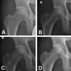
Postoperative radiographs of a 38-year-old patient with avascular necrosis (ARCO stage II) of the right hip. (A) Postoperative x-ray. X-rays of the right hip joint at 6 months (B), 12 months (C), and 24 months (D) after treatment. Over time, the sclerotic and necrotic zones have decreased in size, especially on the lateral side. Despite the extensive defect, the femoral head has neither collapsed nor progressed to an ARCO stage III. After 24 months, the beta-tricalcium phosphate (β-TCP) granula have not undergone complete resorption. Reprinted with permission from Schattauer [54]. ARCO, Association Research Circulation Osseous.
Figure 4
Figure 4
Acellular matrix implantation in a patient with an osteochondral defect of the femoral head and an unstable cartilage defect (ARCO stage III). A trochanteric flip osteotomy [43] was performed to expose the femoral head. (A) Intraoperative view after surgical dislocation, debridement, and anterograde drilling into the sclerotic bone. (B) Aspect of the defect after bone grafting by using cancellous bone from the osteotomy. (C) The cartilaginous portion of the lesion was augmented with an acellular collagen type I hydrogel (CaReS®-OneStep; Arthro Kinetics, Krems, Austria). (D) Schematic illustration of the procedure. ARCO, Association Research Circulation Osseous.

Similar articles

Cited by

References

    1. Mont MA, Hungerford DS. Non-traumatic avascular necrosis of the femoral head. J Bone Joint Surg Am. 1995;77:459–474. - PubMed
    1. Vail TP, Covington DB. In: Osteonecrosis: Etiology, Diagnosis, Treatment. Urbaniak JR, Jones JR, editor. Rosemont, IL: American Academy of Orthopedic Surgeons; 1997. The incidence of osteonecrosis; pp. 43–49.
    1. Lieberman JR, Berry DJ, Mont MA, Aaron RK, Callaghan JJ, Rajadhyaksha AD, Urbaniak JR. Osteonecrosis of the hip: management in the 21st century. Instr Course Lect. 2003;52:337–355. - PubMed
    1. Nöth U, Rackwitz L, Steinert AF, Tuan RS. Cell delivery therapeutics for musculoskeletal regeneration. Adv Drug Deliv Rev. 2010;62:765–783. doi: 10.1016/j.addr.2010.04.004. - DOI - PubMed
    1. Baksh D, Song L, Tuan RS. Adult mesenchymal stem cells: characterization, differentiation, and application in cell and gene therapy. J Cell Mol Med. 2004;8:301–316. doi: 10.1111/j.1582-4934.2004.tb00320.x. - DOI - PMC - PubMed

Publication types

MeSH terms

Substances

LinkOut - more resources