. 2012 Apr 6;287(15):11981-90.
doi: 10.1074/jbc.M111.338525. Epub 2012 Feb 22.

Plant actin-binding protein SCAB1 is dimeric actin cross-linker with atypical pleckstrin homology domain

Affiliations

Plant actin-binding protein SCAB1 is dimeric actin cross-linker with atypical pleckstrin homology domain

Wei Zhang et al. J Biol Chem. .

Abstract

SCAB1 is a novel plant-specific actin-binding protein that binds, bundles, and stabilizes actin filaments and regulates stomatal movement. Here, we dissected the structure and function of SCAB1 by structural and biochemical approaches. We show that SCAB1 is composed of an actin-binding domain, two coiled-coil (CC) domains, and a fused immunoglobulin and pleckstrin homology (Ig-PH) domain. We determined crystal structures for the CC1 and Ig-PH domains at 1.9 and 1.7 Å resolution, respectively. The CC1 domain adopts an antiparallel helical hairpin that further dimerizes into a four-helix bundle. The CC2 domain also mediates dimerization. At least one of the coiled coils is required for actin binding, indicating that SCAB1 is a bivalent actin cross-linker. The key residues required for actin binding were identified. The PH domain lacks a canonical basic phosphoinositide-binding pocket but can bind weakly to inositol phosphates via a basic surface patch, implying the involvement of inositol signaling in SCAB1 regulation. Our results provide novel insights into the functional organization of SCAB1.


Figures
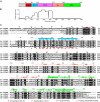
FIGURE 1.
FIGURE 1.
Domain organization and sequence alignment of SCAB1. A, domain diagram of SCAB1. The ABD, CC1, CC2, Ig, and PH domains are labeled. B, prediction of coiled coils by COILS. The prediction was based on the matrix MTK, no positional weighting, and a window of 21. C, multiple-sequence alignment of SCAB1 homologs. Black and gray denote 98 and 80% conservation, respectively, in 33 aligned sequences, among which only SCAB1 homologs from the eudicot A. thaliana (At), the monocot Oryza sativa (Os), the fern Selaginella moellendorffii (Sm), and the moss Physcomitrella patens (Pp) are displayed. The secondary structures observed in the crystal structures of the CC1 and Ig-PH domains are indicated on the top of the alignment. The closed and open circles indicate those residues whose solvent-accessible surface is buried by at least 30 and 10 Å2, respectively, due to CC1 dimerization and are shown on the top and bottom of the alignment for the two subunits of the CC1 dimer. The residues important for F-actin binding are marked with closed squares, and the residues involved in inositol phosphate binding are marked with closed triangles.
FIGURE 2.
FIGURE 2.
Dimeric structure and function of the CC1 domain. A, the omit electron density map is contoured at the 1. 5σ level and superimposed on the final structural model of CC1. B, ribbon representation of the CC1 structure. The two subunits are colored in cyan and magenta. The N and C termini and secondary structures are labeled. C, one subunit of a CC1 dimer is aligned with the nonequivalent subunit of another dimer. D, helical wheel diagram of the CC1 dimer. The αA helices are viewed from the N to C terminus, and the αB helices are viewed from the C to N terminus. Hydrophobic residues are green, Glu and Asp are red, Lys and Arg are blue, and all other residues are black. The lines denote electrostatic interactions. E, interactions stabilizing the CC1 dimeric structure. One subunit (magenta) is depicted as a surface with residues at least 80% conserved colored yellow, whereas the other subunit is shown as ribbons. The interacting residues are shown as sticks. The residues subjected to mutagenesis analysis are encircled. F, F-actin co-sedimentation assay of GFP-tagged SCAB1 NT5 (residues 54–148) and its mutants. NT5 (0.5 μm) was incubated with or without 2 μm preformed F-actin in a 40-μl reaction of 10 mm imidazole (pH 7.0), 75 mm KCl, 1 mm MgCl2, 1 mm EGTA, and 1 mm ATP and subjected to centrifugation at 150,000 × g for 30 min. The proteins in the supernatants (S) and pellets (P) were resolved by SDS-PAGE. G, F-actin co-sedimentation assay of GST-tagged SCAB1 without CC1 (Δ101–148).
FIGURE 3.
FIGURE 3.
Key residues in the ABD. GST-tagged WT SCAB1 and mutants V74A, R75E, L77A, F81A, A88S, and L91A were co-sedimented with F-actin. SCAB1 (0.5 μm) was incubated with or without 2 μm preformed F-actin and subjected to centrifugation at 150,000 × g for 30 min. The proteins in the supernatants (S) and pellets (P) were resolved by SDS-PAGE.
FIGURE 4.
FIGURE 4.
Structure of the Ig-PH domain. A, ribbon representation of the Ig-PH domain. The Ig and PH domains are colored wheat and green, respectively. The secondary structures are labeled. B, the omit electron density map contoured at the 1.5σ level is shown for the malonate-binding site. C, the conservation surface as shown in three orientations. The lower right structure is oriented as in A. The Ig and PH domains are colored wheat and green, respectively. The residues at least 80% conserved are colored yellow. D, interactions between the Ig and PH domains. Hydrogen bonds are denoted by dotted lines.
FIGURE 5.
FIGURE 5.
PH domain of SCAB1 binds inositol phosphates via a low-affinity surface site. A, ITC of SCAB1(272–496) with inositol phosphates Ins(1,4,5)P3, Ins(1,2,3,4,5,6)P6, and Ins(1)P. The curves in the lower panels are the best fit to a one-set-of-sites binding model. The derived dissociation constants (Kd) are indicated. B, ITC of the SCAB1(272–496) mutants R410N, K412N, and K422N with Ins(1,4,5)P3. C, electrostatic potential surface for the PH domain. The surface is colored in blue to red for positively to negatively charged regions. The arrow points to the equivalent high-affinity inositol phosphate (IP)-binding pocket, which is not negatively charged in SCAB1. D, interaction between the SCAB1 PH domain and malonate. The dotted lines denote hydrogen bonds. E, structure of the phospholipase Cδ1 (PLC-δ) PH domain in complex with Ins(1,4,5)P3 (Protein Data Bank code 1MAI) aligned with the SCAB1 PH domain structure. F, structure of the β-spectrin PH domain in complex with Ins(1,4,5)P3 (Protein Data Bank code 1BTN) aligned with the SCAB1 PH domain structure.
FIGURE 6.
FIGURE 6.
Full-length SCAB1 is a dimer. A, the C-terminal part of CC2 mediates dimerization. The Ig-PH domain alone (residues 272–496) and the Ig-PH domain fused to part of CC2 (residues 239–496) were analyzed using a Superdex 200 10/300 column. The normalized absorbance at 215 nm is plotted against the elution volume. The elution positions of the calibration standards BSA (67 kDa), chymotrypsinogen A (25 kDa), and lysozyme (14.7 kDa) are marked. B, analytic ultracentrifuge sedimentation equilibrium analysis of full-length SCAB1. The curves are the best global fit of the three profiles to the single-species model, yielding a molecular mass of 102.8 ± 1.0 kDa.
FIGURE 7.
FIGURE 7.
Model depicting a SCAB1 dimer that cross-links AFs. SCAB1 dimerizes via its CC1 and CC2 domains. CC2 is shown as parallel coils but may adopt a more complex structure. The dual ABD in the SCAB1 dimer simultaneously contact two adjacent AFs.

Similar articles

Cited by

References

    1. Pollard T. D., Cooper J. A. (2009) Actin, a central player in cell shape and movement. Science 326, 1208–1212 - PMC - PubMed
    1. Blanchoin L., Boujemaa-Paterski R., Henty J. L., Khurana P., Staiger C. J. (2010) Actin dynamics in plant cells: a team effort from multiple proteins orchestrates this very fast-paced game. Curr. Opin. Plant Biol. 13, 714–723 - PubMed
    1. Hussey P. J., Ketelaar T., Deeks M. J. (2006) Control of the actin cytoskeleton in plant cell growth. Annu. Rev. Plant Biol. 57, 109–125 - PubMed
    1. Higaki T., Kojo K. H., Hasezawa S. (2010) Critical role of actin bundling in plant cell morphogenesis. Plant Signal. Behav. 5, 484–488 - PMC - PubMed
    1. Thomas C., Tholl S., Moes D., Dieterle M., Papuga J., Moreau F., Steinmetz A. (2009) Actin bundling in plants. Cell Motil. Cytoskeleton 66, 940–957 - PubMed

Publication types

MeSH terms