. 2012 Apr 20;287(17):14023-39.
doi: 10.1074/jbc.M111.318352. Epub 2012 Feb 22.

Direct evidence of generation and accumulation of β-sheet-rich prion protein in scrapie-infected neuroblastoma cells with human IgG1 antibody specific for β-form prion protein

Affiliations

Direct evidence of generation and accumulation of β-sheet-rich prion protein in scrapie-infected neuroblastoma cells with human IgG1 antibody specific for β-form prion protein

Toshiya Kubota et al. J Biol Chem. .

Abstract

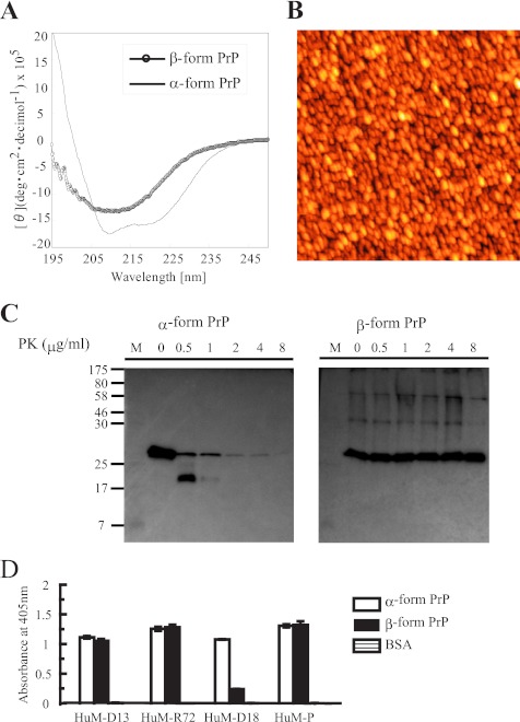
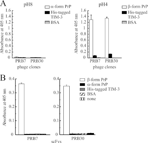
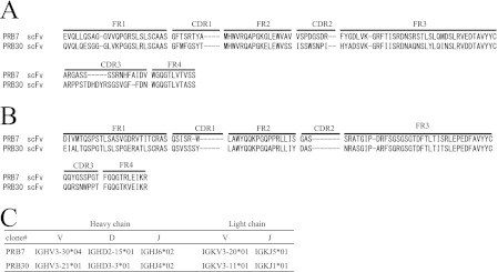
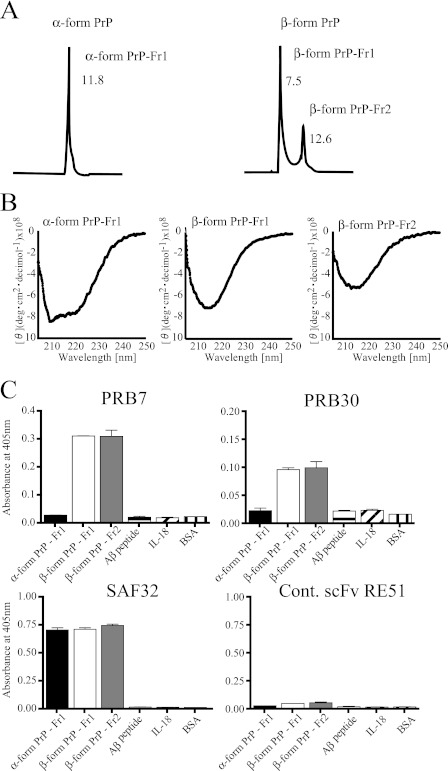
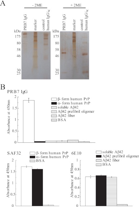
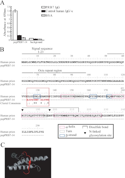
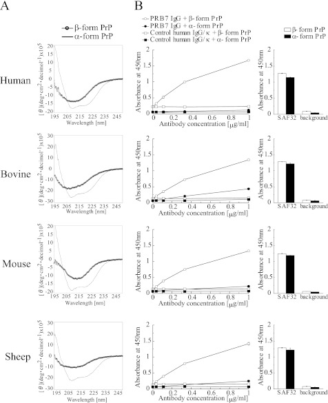
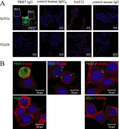
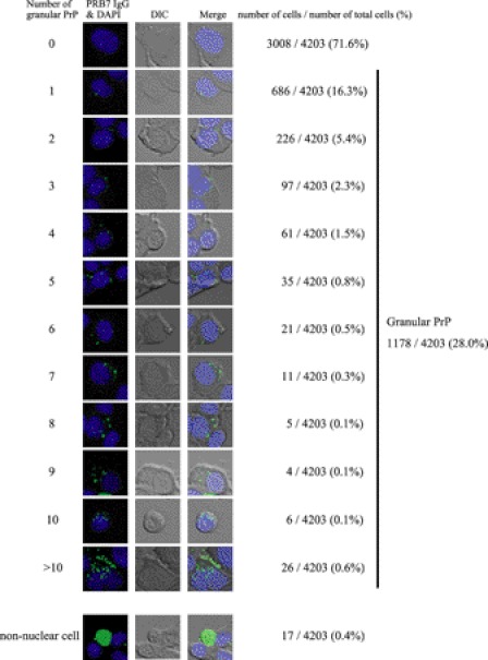
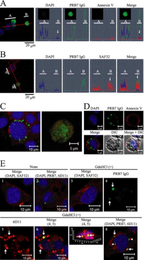
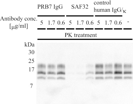
We prepared β-sheet-rich recombinant full-length prion protein (β-form PrP) (Jackson, G. S., Hosszu, L. L., Power, A., Hill, A. F., Kenney, J., Saibil, H., Craven, C. J., Waltho, J. P., Clarke, A. R., and Collinge, J. (1999) Science 283, 1935-1937). Using this β-form PrP and a human single chain Fv-displaying phage library, we have established a human IgG1 antibody specific to β-form but not α-form PrP, PRB7 IgG. When prion-infected ScN2a cells were cultured with PRB7 IgG, they generated and accumulated PRB7-binding granules in the cytoplasm with time, consequently becoming apoptotic cells bearing very large PRB7-bound aggregates. The SAF32 antibody recognizing the N-terminal octarepeat region of full-length PrP stained distinct granules in these cells as determined by confocal laser microscopy observation. When the accumulation of proteinase K-resistant PrP was examined in prion-infected ScN2a cells cultured in the presence of PRB7 IgG or SAF32, it was strongly inhibited by SAF32 but not at all by PRB7 IgG. Thus, we demonstrated direct evidence of the generation and accumulation of β-sheet-rich PrP in ScN2a cells de novo. These results suggest first that PRB7-bound PrP is not responsible for the accumulation of β-form PrP aggregates, which are rather an end product resulting in the triggering of apoptotic cell death, and second that SAF32-bound PrP lacking the PRB7-recognizing β-form may represent so-called PrP(Sc) with prion propagation activity. PRB7 is the first human antibody specific to β-form PrP and has become a powerful tool for the characterization of the biochemical nature of prion and its pathology.


Figures

FIGURE 1.
FIGURE 1.
Characterization of human full-length prion protein refolded in vitro. Refolding was performed according to the protocol of Jackson et al. (12). A, CD spectra for the protein refolded at pH 8.0 (line; α-form PrP) and the protein refolded at pH 4.0 (open circle; β-form PrP). A double minimum at 222 and 208 nm or a minimum at ∼215 nm is characteristic of an α-helical structure or β-sheet structure, respectively (26). B, atomic force microscopy image of β-form PrP. C, limited PK digestion of β-form PrP. α-Form PrP or β-form PrP (1 μg) was incubated with varying concentrations (0–8 μg/ml) of PK for 30 min at 37 °C. PrP was detected by immunoblot analysis with the 6D11 antibody. D, reactivity of α-form PrP or β-form PrP with HuM anti-PrP antibodies (–19). Region 133–157 recognized by the anti-PrP Fab antibody (D18) is cryptic in β-PrP. Data represent the mean values ±S.D. deg, degrees.
FIGURE 2.
FIGURE 2.
Binding specificity of β-form PrP-selected scFv phage clones by ELISA. A, phage clones (PRB7 or PRB30; 5 × 10¹¹ transforming units/40 μl) were incubated with the indicated antigens (200 ng/40 μl) in a pH 8.0 or 4.0 folding buffer for 1 h and then added to wells coated with an anti-His tag mAb. The bound phage was detected using a biotinylated anti-M13 monoclonal antibody (1:1000) followed by the addition of alkaline phosphatase-conjugated streptavidin (1:1000) as described under “Experimental Procedures.” His-tagged TIM-3 or BSA was used as a control protein. B, soluble PRB7 or PRB30 scFv antibody (1 μg/200 μl) was added to the indicated antigen-coated ELISA plates (200 ng/40 μl/well) using pH 7.3 binding buffer. The bound scFv antibodies were detected using a biotinylated anti-E tag monoclonal antibody (1:1000) followed by the addition of alkaline phosphatase-conjugated streptavidin (1:1000). Data represent the mean values ±S.D.
FIGURE 3.
FIGURE 3.
Amino acid sequence analysis of identified scFv specific to β-PrP. A, Vh domains. B, Vl domains. C, the germ line gene usage of anti-β-PrP scFv. Given are the names of the gene segments according to the IMGT database. *, other possibilities: IGHJ5*01 and IGHJ4*01 (highest number of consecutive identical nucleotides) or IGHJ4*01 (shorter alignment but highest percentage of identity).
FIGURE 4.
FIGURE 4.
PRB7 and PRB30 scFv recognize β-form PrP monomer and oligomer fractionated by SEC. A, SEC profile of α-form PrP (left) and β-form PrP (right). α-Form PrP or β-form PrP was eluted with a running buffer in the presence of 1 m urea at pH 5.5 or a running buffer in the presence of 1 m urea at pH 4.0, respectively. B, CD spectra of SEC-fractionated samples. The CD signals at wavelengths below 200 nm (not shown) are distorted as a result of absorption by urea. C, the scFv antibodies were added to wells coated with SEC-fractionated samples. PRB7 or PRB30 scFv antibody recognized β-form PrP-Fr1 and β-form PrP-Fr2 but not α-form PrP-Fr1, amyloid β42 peptide, or IL-18. Unrelated scFv (RE51) showed no binding activity to any of these proteins. Data represent the mean values ±S.D. Cont., control; deg, degrees.
FIGURE 5.
FIGURE 5.
Characterization of PRB7 IgG antibody. A, SDS-PAGE of PRB7 IgG. Protein A column-purified PRB7 IgG (300 ng) was separated by 10% SDS-PAGE under reducing or non-reducing condition and detected by silver staining. B, PrP conformers (50 ng/50 μl/well) were detected by PRB7 IgG or SAF32 (0.5 μg/ml). Aβ42 conformers (50 ng/50 μl/well) were tested for binding reactivity to PRB7 IgG or 6E10 (mouse anti-Aβ42 antibody) as described under “Experimental Procedures.” 2ME, 2-mercaptoethanol. Data represent the mean values ±S.D.
FIGURE 6.
FIGURE 6.
Epitope mapping of PRB7 IgG. A, peptide phage clones (50 μl/well of 1 × 10¹¹ virions/ml) were incubated with PRB7 IgG (50 ng/50 μl/well). The bound peptide phage clones were detected using a biotinylated anti-M13 monoclonal antibody (1:2000) followed by HRP-conjugated streptavidin (1:2000). The pepPRB7-14 was selected from a Ph.D.-12 peptide phage library. Data represent the mean values ±S.D. B, amino acid sequence homology between the full-length human PrP and pepPRB7-14 motif (red) was searched by ClustalW. The matching region (amino acids 124–135) is depicted in the red box. The secondary structures are indicated by explanatory marks as shown in the black box. C, computer simulation model of human α-form PrP (Protein Data Bank code 1QM0). The matching region (amino acids 124–135) containing a highly homologous region (amino acids 128–132) is depicted as a red ribbon. PRB7 IgG does not recognize this structure of α-form PrP.
FIGURE 7.
FIGURE 7.
Binding specificity of PRB7 IgG by ELISA analysis. A, CD spectra of α-form PrP and β-form PrP. Recombinant PrP was refolded as described under “Experimental Procedures.” B, ELISA of PRB7 IgG antibodies binding to PrP. Left panel, PRB7 IgG (ranging from 0 to 1 μg/ml) was incubated with PrP-coated plates (100 ng/50 μl/well). Normal human IgG was used as a control. The secondary antibody was HRP-conjugated anti-human IgG (1:5000 dilution). Right panel, SAF32 (0.1 μg/ml) equally binds to both α-form PrP and β-form PrP (100 ng/50 μl/well). The background was determined using HRP-conjugated anti-human IgG alone. Data represent the mean values ±S.D. deg, degrees.
FIGURE 8.
FIGURE 8.
β-Form PrPs are generated and accumulate de novo in prion-infected ScN2a cells. A, ScN2a or N2a58 cells were cultured in the absence or presence of PRB7 IgG, SAF32, control human IgG/κ, or control mouse IgG (5 μg/ml) in 10% FBS, Opti-MEM. Three days later, cells were fixed with paraformaldehyde and permeated for staining with Alexa Fluor 488-labeled anti-human IgG (green), Alexa Fluor 546-labeled anti-mouse IgG (red), or DAPI (blue) as described under “Experimental Procedures.” Each box indicates a representative staining pattern. Box 1, Box 2, and Box 3 show Large/many granules, a few tiny granules, or no granules stained with PRB7 IgG, respectively. B, ScN2a cells cultured in the presence of PRB7 IgG or control human IgG/κ (hIgG) (green) as described in A were fixed with methanol and stained with anti-β-actin antibody (red), anti-β-tubulin antibody (red), or DAPI (blue) as described under “Experimental Procedures.”
FIGURE 9.
FIGURE 9.
Distribution pattern of PRB7 IgG-positive granules in ScN2a cells cultured with PRB7 IgG. Cells were stained with PRB7 IgG or DAPI as described in Fig. 8. A total of 4203 cells were classified by the number of granules in the cytoplasm (positive cells/total cells observed). DIC, differential interference contrast.
FIGURE 10.
FIGURE 10.
ScN2a cells exhibit PRB7 IgG-positive aggregates under apoptosis. Cells were stained with PRB7 IgG (green), SAF32 (red), or DAPI (blue) as described in Fig. 8. A, ScN2a cells with very large aggregates in the cytoplasm underwent apoptosis. ScN2a cells cultured with PRB7 IgG were incubated with Annexin V (red) followed by fixing, permeation, and staining with anti-human IgG or DAPI as described under “Experimental Procedures.” Orthogonal projections of serial confocal sections are shown by histograms alongside one z-section taken from the middle of the cell (as indicated by the red guidelines). White bar A or B indicates the range of the guideline. The white arrow in the B range indicates the pattern of a huge aggregate-bearing ScN2a cell. Scale bar, 20 μm. B, very large aggregates in the cytoplasm are PrP. ScN2a cells cultured with PRB7 IgG were fixed, permeated, and stained with SAF32 followed by secondary antibody staining with Alexa Fluor 488-labeled anti-human IgG or Alexa Fluor 546-labeled anti-mouse IgG. Orthogonal projections of serial confocal sections are shown by histograms alongside one z-section taken from the middle of the cell (as indicated by the red guidelines). White bar A or B indicates the range of the guideline. The white arrow in the B range indicates the pattern of a huge aggregate-bearing ScN2a cell. C, PRB7 IgG-positive images do not merge with SAF32-positive images. Left image, prion-infected ScN2a cells containing a number of small PRB7 IgG-bound granules. SAF32 stained the entire region of cytoplasm except PRB7 IgG-bound granules. Right image, prion-infected ScN2a cells containing large tangled fibrillary aggregates stained with PRB7 IgG. A number of tiny granules were stained with SAF32 independently of PRB7 IgG-positive aggregates. D, ScN2a cells with a number of PRB7 IgG-positive granules around nuclei underwent apoptosis. ScN2a cells cultured with PRB7 IgG were incubated with Annexin V followed by fixing, permeation, and staining with anti-human IgG or DAPI as described in A. E, ScN2a cells cultured with Alexa Fluor 488-PRB7 IgG were fixed, permeated, treated with (+) or without (−) 6 m GdnHCl, and stained with 6D11 (epitope 93–109 of PrP (32)). Panel 1 is an image merged with DAPI and SAF32 staining of non-treated ScN2a cells. Panel 2 is an image merged with DAPI, PRB7 IgG, and 6D11 staining of non-treated ScN2a cells. Panels 3–8 show the images of 6 m GdnHCl-treated ScN2a cells. Panels 4–7 show images of an identical cell. Panel 3 is an image merged with DAPI and SAF32 staining. Panel 4 is an image stained with Alexa Fluor 488-PRB7 IgG. Panel 5 is an image stained with 6D11. Panel 6 is an image merged with PRB7 IgG and 6D11 staining. Panel 7 is a three-dimensional image of panel 6. Panel 8 shows a cell containing heterogeneous PRB7 IgG-bound aggregates merged with (white arrow) or without (white arrowhead) 6D11 staining in the presence of numerous tiny aggregates stained with 6D11 in the cytoplasm. Staining was performed as described under “Experimental Procedures.” DIC, differential interference contrast.
FIGURE 11.
FIGURE 11.
Effect of PRB7 IgG on accumulation of PrPres. ScN2a cells were cultured with PRB7 IgG, SAF32, or control human IgG/κ antibodies ranging from 0 to 5 μg/ml for 4 days at 37 °C. In the case of “−”, ScN2a was cultured without antibody for 4 days at 37 °C. Cell lysates were treated with PK (final concentration (conc.), 20 μg/ml) as described under “Experimental Procedures.” Lysates were separated on a 16.5% Tricine-polyacrylamide gel and immunoblotted with 6D11 antibody followed by HRP-conjugated anti-goat mouse IgG as described under “Experimental Procedures.”

Similar articles

Cited by

References

    1. Prusiner S. B. (1998) Prions. Proc. Natl. Acad. Sci. U.S.A. 95, 13363–13383 - PMC - PubMed
    1. Rogers M., Yehiely F., Scott M., Prusiner S. B. (1993) Conversion of truncated and elongated prion proteins into the scrapie isoform in cultured cells. Proc. Natl. Acad. Sci. U.S.A. 90, 3182–3186 - PMC - PubMed
    1. Pan K. M., Baldwin M., Nguyen J., Gasset M., Serban A., Groth D., Mehlhorn I., Huang Z., Fletterick R. J., Cohen F. E.(1993) Conversion of α-helices into β-sheets features in the formation of the scrapie prion proteins. Proc. Natl. Acad. Sci. U.S.A. 90, 10962–10966 - PMC - PubMed
    1. Prusiner S. B., McKinley M. P., Bowman K. A., Bolton D. C., Bendheim P. E., Groth D. F., Glenner G. G. (1983) Scrapie prions aggregate to form amyloid-like birefringent rods. Cell 35, 349–358 - PubMed
    1. Turk E., Teplow D. B., Hood L. E., Prusiner S. B. (1988) Purification and properties of the cellular and scrapie hamster prion proteins. Eur. J. Biochem. 176, 21–30 - PubMed

Publication types