. 2012 May;86(9):5264-77.
doi: 10.1128/JVI.06913-11. Epub 2012 Feb 22.

Herpes simplex virus 1 VP22 regulates translocation of multiple viral and cellular proteins and promotes neurovirulence

Affiliations

Herpes simplex virus 1 VP22 regulates translocation of multiple viral and cellular proteins and promotes neurovirulence

Michiko Tanaka et al. J Virol. 2012 May.

Abstract

Herpes simplex virus 1 (HSV-1) protein VP22, encoded by the UL49 gene, is a major virion tegument protein. In the present study, we showed that VP22 was required for efficient redistribution of viral proteins VP16, VP26, ICP0, ICP4, and ICP27 and of cellular protein Hsc-70 to the cytoplasm of infected cells. We found that two dileucine motifs in VP22, at amino acids 235 and 236 and amino acids 251 and 252, were necessary for VP22 regulation of the proper cytoplasmic localization of these viral and cellular proteins. The dileucine motifs were also required for proper cytoplasmic localization of VP22 itself and for optimal expression of viral proteins VP16, VP22, ICP0, UL41, and glycoprotein B. Interestingly, a recombinant mutant virus with alanines substituted for the dileucines at amino acids 251 and 252 had a 50% lethal dose (LD(50)) for neurovirulence in mice following intracerebral inoculation about 10(3)-fold lower than the LD(50) of the repaired virus. Furthermore, the replication and spread of this mutant virus in the brains of mice following intracerebral inoculation were significantly impaired relative to those of the repaired virus. The ability of VP22 to regulate the localization and expression of various viral and cellular proteins, as shown in this study, was correlated with an increase in viral replication and neurovirulence in the experimental murine model. Thus, HSV-1 VP22 is a significant neurovirulence factor in vivo.


Figures
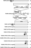
Fig 1
Fig 1
Schematic diagrams of the genome structures of wild-type YK304 and the relevant domains of the recombinant viruses used in this study. Diagram 1, the YK304 genome carrying a bacmid (BAC) in the intergenic region between UL3 and UL4. Diagram 2, domains carrying the UL48 to UL50 open reading frames. Diagram 3, the UL49 gene, encoding VP22. Diagrams 4 to 12, recombinant viruses with mutations in the VP22 gene. Diagram 13, domains carrying the UL40 to UL42 open reading frames. Diagram 14, the UL41 gene, encoding VHS. Diagram 15, recombinant virus with a mutation in the UL41 gene.
Fig 2
Fig 2
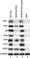
Immunoblots of electrophoretically separated lysates of Vero cells that were either mock infected (lane 4) or infected with wild-type HSV-1(F) (lane 1), YK451 (ΔVP22) (lane 2), or YK452 (ΔVP22-repair) (lane 3) at an MOI of 1. Infected Vero cells were harvested at 20 h postinfection and were immunoblotted with an antibody to VP22, VP16, gB, ICP0, ICP4, UL41, ICP27, or α-tubulin.
Fig 3
Fig 3
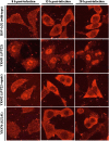
Digital confocal microscope images showing the localization of VP16. Vero cells were infected with wild-type HSV-1(F), YK451 (ΔVP22), YK452 (ΔVP22-repair), or YK476 (ΔUL41) at an MOI of 1. Infected Vero cells were fixed at the indicated times postinfection, permeabilized, stained with an anti-VP16 antibody, and examined by confocal microscopy.
Fig 4
Fig 4
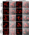
Digital confocal microscope images showing the localization of VP16 in Vero cells infected with wild-type HSV-1(F), YK451 (ΔVP22), YK453 (VP22LL235AA), YK454 (VP22LL235AA-repair), YK455 (VP22LL251AA), or YK456 (VP22LL251AA-repair) at an MOI of 1 for 15 h. Infected Vero cells were fixed, permeabilized, stained with an anti-VP16 antibody, and examined by confocal microscopy.
Fig 5
Fig 5
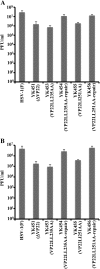
Effects of the ΔVP22, VP22LL235AA, and VP22LL251AA mutations on the production of infectious virus. Vero cells were infected with wild-type HSV-1(F), YK451 (ΔVP22), YK453 (VP22LL235AA), YK454 (VP22LL235AA-repair), YK455 (VP22LL251AA), or YK456 (VP22LL251AA-repair) at an MOI of 1 (A) or 0.01 (B). Total virus from the cell culture supernatants and infected cells was harvested at 24 h postinfection and was assayed on Vero cells. Data are means and standard errors for three independent experiments.
Fig 6
Fig 6
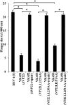
Effects of the ΔVP22, VP22LL235AA, and VP22LL251AA mutations on the production of plaques. Vero cells were infected with wild-type HSV-1(F), YK451 (ΔVP22), YK452 (ΔVP22-repair), YK453 (VP22LL235AA), YK454 (VP22LL235AA-repair), YK455 (VP22LL251AA), or YK456 (VP22LL251AA-repair) at an MOI of 0.0001 under plaque assay conditions. The diameters of 20 single plaques for each of the recombinant viruses were determined 2 days after infection. The data shown are means and standard errors. Asterisks indicate significant differences (*, P < 1 × 10⁻⁸) by a two-tailed Student t test.
Fig 7
Fig 7
Digital confocal microscope images showing the localization of VP22. Vero cells were infected with wild-type HSV-1(F), YK453 (VP22LL235AA), YK454 (VP22LL235AA-repair), YK455 (VP22LL251AA), or YK456 (VP22LL251AA-repair) at an MOI of 1 for 15 h. Infected Vero cells were fixed at the indicated times postinfection, permeabilized, stained with an anti-VP22 antibody, and examined by confocal microscopy. The left and right columns at each time point show VP22 fluorescence and the simultaneous acquisition of VP22 fluorescence and digital interference contrast, respectively.
Fig 8
Fig 8
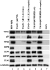
Immunoblots of electrophoretically separated lysates of Vero cells that were either mock infected (lane 7) or infected with wild-type HSV-1(F) (lane 1), YK451 (ΔVP22) (lane 2), YK453 (VP22LL235AA) (lane 3), YK454 (VP22LL235-repair) (lane 4), YK455 (VP22LL251AA) (lane 5), or YK456 (VP22LL251AA-repair) (lane 6) at an MOI of 1. Infected Vero cells were harvested 20 h postinfection and were immunoblotted with an antibody to VP16, gB, VP22, ICP0, ICP4, ICP27, UL41, or α-tubulin.
Fig 9
Fig 9
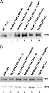
(A) Immunoblots of electrophoretically separated virions of wild-type HSV-1(F) (lane 1), YK453 (VP22LL235AA) (lane 2), YK454 (VP22LL235AA-repair) (lane 3), YK456 (VP22LL251AA-repair) (lane 4), YK455 (VP22LL251AA) (lane 5), and YK476 (ΔUL41) (lane 6) purified by sucrose gradient centrifugation and blotted with an antibody to VP22. (B) Immunoblots of electrophoretically separated virions of wild-type HSV-1(F) (lane 1), YK453 (VP22LL235AA) (lane 2), YK454 (VP22LL235AA-repair) (lane 3), YK455 (VP22LL251AA) (lane 4), YK456 (VP22LL251AA-repair) (lane 5), and YK476 (ΔUL41) (lane 6) purified by sucrose gradient centrifugation and blotted with an antibody to VP5 or VP16.
Fig 10
Fig 10
Digital confocal microscope images showing the localization of ICP4 and ICP8 in Vero cells infected with wild-type HSV-1(F), YK451 (ΔVP22), YK452 (ΔVP22-repair), YK455 (VP22LL251AA), YK456 (VP22LL251AA-repair), or YK476 (ΔUL41) at an MOI of 1. Infected Vero cells were fixed at the indicated times postinfection, permeabilized, stained with an antibody to ICP4 or ICP8, and examined by confocal microscopy. Left and right columns at each time point show protein fluorescence and simultaneous acquisition of protein fluorescence and digital interference contrast, respectively.
Fig 11
Fig 11
Digital confocal microscope images showing the localization of ICP27 and VP26 in Vero cells infected with wild-type HSV-1(F), YK451 (ΔVP22), YK452 (ΔVP22-repair), YK455 (VP22LL251AA), YK456 (VP22LL251AA-repair), or YK476 (ΔUL41) at an MOI of 1. Infected Vero cells were fixed at the indicated times postinfection, permeabilized, stained with an antibody to ICP27 or VP26, and examined by confocal microscopy. Left and right columns at each time point show protein fluorescence and simultaneous acquisition of protein fluorescence and digital interference contrast, respectively.
Fig 12
Fig 12
Digital confocal microscope images showing the localization of ICP0 in Vero cells infected with wild-type HSV-1(F), YK451 (ΔVP22), YK452 (ΔVP22-repair), YK455 (VP22LL251AA), YK456 (VP22LL251AA-repair), or YK476 (ΔUL41) at an MOI of 1. Infected Vero cells were fixed at the indicated times postinfection, permeabilized, stained with an antibody to ICP0, and examined by confocal microscopy. Left and right columns at each time point show protein fluorescence and simultaneous acquisition of protein fluorescence and digital interference contrast, respectively.
Fig 13
Fig 13
Digital confocal microscope images showing the localization of Hsc70 and AIF in normal Vero cells (A) and in Vero cells infected with wild-type HSV-1(F), YK451 (ΔVP22), YK452 (ΔVP22-repair), YK455 (VP22LL251AA), YK456 (VP22LL251AA-repair), or YK476 (ΔUL41) at an MOI of 1 (B). Infected Vero cells were fixed at the indicated times postinfection, permeabilized, stained with an antibody to Hsc70 or AIF, and examined by confocal microscopy. Left and right columns at each time point show protein fluorescence and simultaneous acquisition of protein fluorescence and digital interference contrast, respectively.
Fig 14
Fig 14
(A) LD50 values of YK455 (VP22LL251AA) and YK456 (VP22LL251AA-repair) in mice following intracerebral inoculation. Three-week-old female mice were inoculated intracerebrally with serial 10-fold dilutions of each virus in groups of 6 per dilution and were monitored for 14 days. LD50 values were determined by the Behrens-Karber method. (B) Virus titers in the brains of mice following intracerebral inoculation. Seventeen 3-week-old female mice were inoculated intracerebrally with 102 PFU of each virus. At 3 days postinfection, the brains of infected mice were harvested, and virus titers were determined by standard plaque assays on Vero cells. The data shown are means and standard errors. A statistically significant difference in virus titer between mice infected with YK455 (VP22LL251AA) and those infected with YK456 (VP22LL251AA-repair) was noted (*, P < 0.005).
Fig 15
Fig 15
Histopathological features of the brains of mice following intracerebral inoculation. Five 3-week-old female mice were inoculated intracerebrally with 102 PFU of YK455 (VP22LL251AA) (A, B, and C) or YK456 (VP22LL251AA-repair) (D, E, and F). At 5 days postinfection, the brains of infected mice were harvested, sectioned, and stained with hematoxylin and eosin (A and D) or with an antibody to HSV-1 antigens (B, C, E, and F). Panels C and F show magnified images of the regions indicated in panels B and E, respectively. Representative images are shown. Bars, 50 μm.

Similar articles

Cited by

References

    1. Asai R, Ohno T, Kato A, Kawaguchi Y. 2007. Identification of proteins directly phosphorylated by UL13 protein kinase from herpes simplex virus 1. Microbes Infect. 9:1434–1438 - PubMed
    1. Bastian TW, Livingston CM, Weller SK, Rice SA. 2010. Herpes simplex virus type 1 immediate-early protein ICP22 is required for VICE domain formation during productive viral infection. J. Virol. 84:2384–2394 - PMC - PubMed
    1. Blaho JA, Mitchell C, Roizman B. 1994. An amino acid sequence shared by the herpes simplex virus 1 alpha regulatory proteins 0, 4, 22, and 27 predicts the nucleotidylylation of the UL21, UL31, UL47, and UL49 gene products. J. Biol. Chem. 269:17401–17410 - PubMed
    1. Brignati MJ, Loomis JS, Wills JW, Courtney RJ. 2003. Membrane association of VP22, a herpes simplex virus type 1 tegument protein. J. Virol. 77:4888–4898 - PMC - PubMed
    1. Burch AD, Weller SK. 2004. Nuclear sequestration of cellular chaperone and proteasomal machinery during herpes simplex virus type 1 infection. J. Virol. 78:7175–7185 - PMC - PubMed

Publication types

MeSH terms