

Roles of E4orf6 and VA I RNA in adenovirus-mediated stimulation of human parvovirus B19 DNA replication and structural gene expression
- PMID: 22357277
- PMCID: PMC3347340
- DOI: 10.1128/JVI.06991-11
Roles of E4orf6 and VA I RNA in adenovirus-mediated stimulation of human parvovirus B19 DNA replication and structural gene expression
Abstract
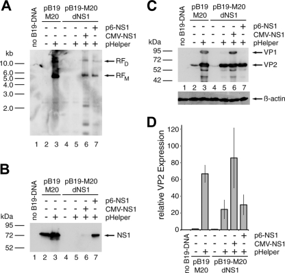
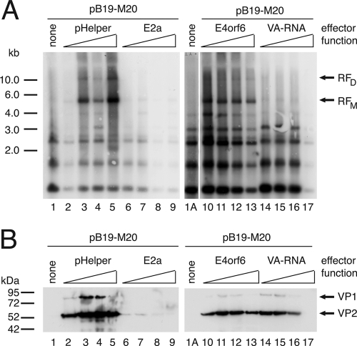
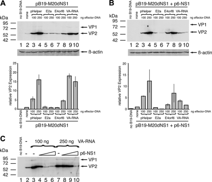
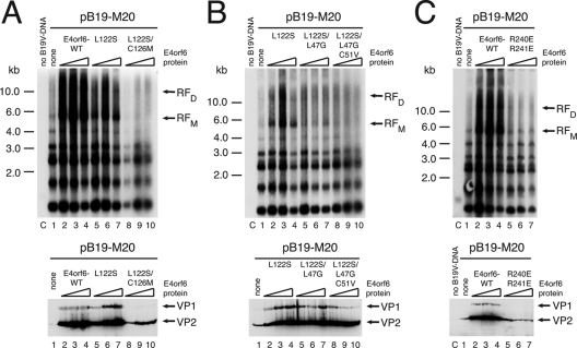
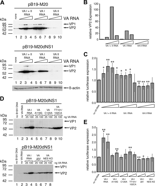
Despite its very narrow tropism for erythroid progenitor cells, human parvovirus B19 (B19V) has recently been shown to replicate and form infectious progeny virus in 293 cells in the presence of early adenoviral functions provided either by infection with adenovirus type 5 or by addition of the pHelper plasmid encoding the E2a, E4orf6, and VA RNA functions. In the present study we dissected the individual influence of these functions on B19V genome replication and expression of structural proteins VP1 and VP2. We show that, in the presence of the constitutively expressed E1A and E1B, E4orf6 alone is able to promote B19V DNA replication, resulting in a concomitant increase in VP expression levels. The stimulatory effects of E4orf6 require the integrity of the BC box motifs, which target cellular proteins such as p53 and the Mre11 DNA repair complex for proteosomal degradation through formation of an E3 ubiquitin ligase complex with E1B. VA RNA also strongly induces VP expression but, in contrast to E4orf6, in a replication-independent manner. This stimulation could be attributed exclusively to the VA I RNA transcript and does not involve major activating effects at the level of the B19V p6 promoter, but the nucleotide residues required for the well-defined pathway of VA I RNA mediated stimulation of translation through functional inactivation of protein kinase R. These data show that the cellular pathways regulating B19V replication may be very similar to those governing the productive cycle of the helper-dependent parvoviruses, the adeno-associated viruses.
Figures
Similar articles
-
The genome of human parvovirus b19 can replicate in nonpermissive cells with the help of adenovirus genes and produces infectious virus.J Virol. 2009 Sep;83(18):9541-53. doi: 10.1128/JVI.00702-09. Epub 2009 Jul 8. J Virol. 2009. PMID: 19587029 Free PMC article.
-
Identification of integrin alpha3 as a new substrate of the adenovirus E4orf6/E1B 55-kilodalton E3 ubiquitin ligase complex.J Virol. 2009 Jun;83(11):5329-38. doi: 10.1128/JVI.00089-09. Epub 2009 Mar 18. J Virol. 2009. PMID: 19297475 Free PMC article.
-
Both BC-box motifs of adenovirus protein E4orf6 are required to efficiently assemble an E3 ligase complex that degrades p53.Mol Cell Biol. 2004 Nov;24(21):9619-29. doi: 10.1128/MCB.24.21.9619-9629.2004. Mol Cell Biol. 2004. PMID: 15485928 Free PMC article.
-
Recent Advances in Replication and Infection of Human Parvovirus B19.Front Cell Infect Microbiol. 2018 Jun 5;8:166. doi: 10.3389/fcimb.2018.00166. eCollection 2018. Front Cell Infect Microbiol. 2018. PMID: 29922597 Free PMC article. Review.
-
Persistent parvovirus B19 infection in non-erythroid tissues: possible role in the inflammatory and disease process.Virus Res. 2014 Sep 22;190:8-16. doi: 10.1016/j.virusres.2014.06.017. Epub 2014 Jul 3. Virus Res. 2014. PMID: 24998884 Review.
Cited by
-
High Prevalence of Infectious Adeno-associated Virus (AAV) in Human Peripheral Blood Mononuclear Cells Indicative of T Lymphocytes as Sites of AAV Persistence.J Virol. 2017 Jan 31;91(4):e02137-16. doi: 10.1128/JVI.02137-16. Print 2017 Feb 15. J Virol. 2017. PMID: 27928011 Free PMC article.
-
Possible involvement of miRNAs in tropism of Parvovirus B19.Mol Biol Rep. 2016 Mar;43(3):175-81. doi: 10.1007/s11033-016-3952-8. Epub 2016 Feb 15. Mol Biol Rep. 2016. PMID: 26878856
-
Comprehensive Small RNA-Seq of Adeno-Associated Virus (AAV)-Infected Human Cells Detects Patterns of Novel, Non-Coding AAV RNAs in the Absence of Cellular miRNA Regulation.PLoS One. 2016 Sep 9;11(9):e0161454. doi: 10.1371/journal.pone.0161454. eCollection 2016. PLoS One. 2016. PMID: 27611072 Free PMC article.
-
Differential adeno-associated virus serotype-specific interaction patterns with synthetic heparins and other glycans.J Virol. 2014 Mar;88(5):2991-3003. doi: 10.1128/JVI.03371-13. Epub 2013 Dec 26. J Virol. 2014. PMID: 24371066 Free PMC article.
-
Human parvovirus B19: a mechanistic overview of infection and DNA replication.Future Virol. 2015;10(2):155-167. doi: 10.2217/fvl.14.103. Future Virol. 2015. PMID: 26097496 Free PMC article.
References
Publication types
MeSH terms
Substances
LinkOut - more resources
Full Text Sources
Other Literature Sources
Molecular Biology Databases
Research Materials
Miscellaneous
