. 2012 May;86(9):5341-51.
doi: 10.1128/JVI.06243-11. Epub 2012 Feb 22.

High-risk human papillomavirus E5 oncoprotein displays channel-forming activity sensitive to small-molecule inhibitors

Affiliations

High-risk human papillomavirus E5 oncoprotein displays channel-forming activity sensitive to small-molecule inhibitors

Laura F Wetherill et al. J Virol. 2012 May.

Abstract

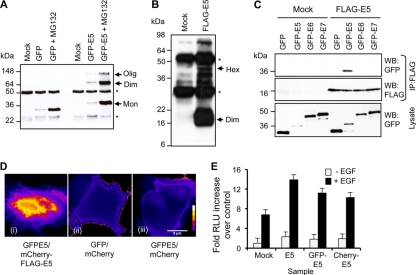
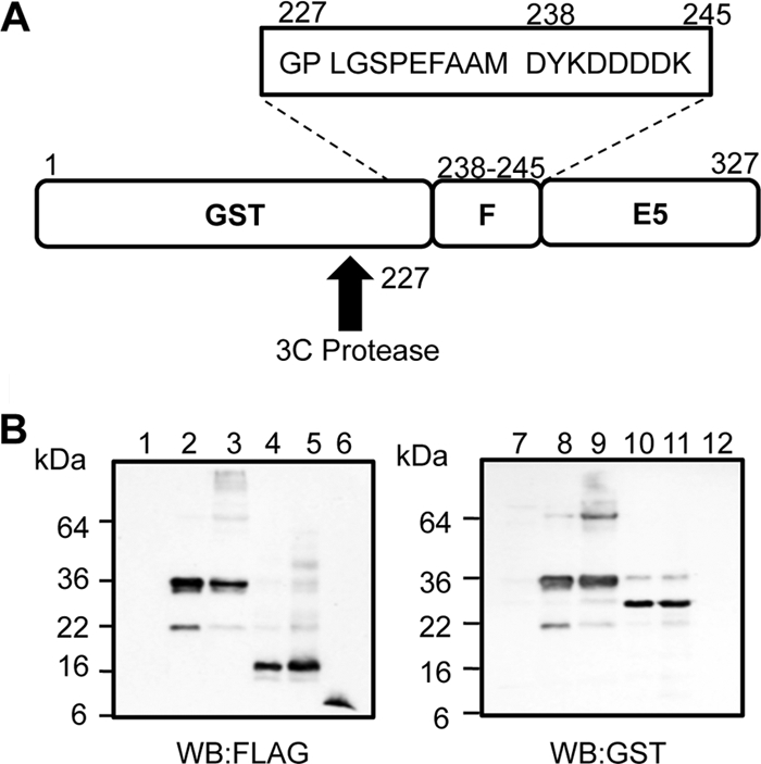
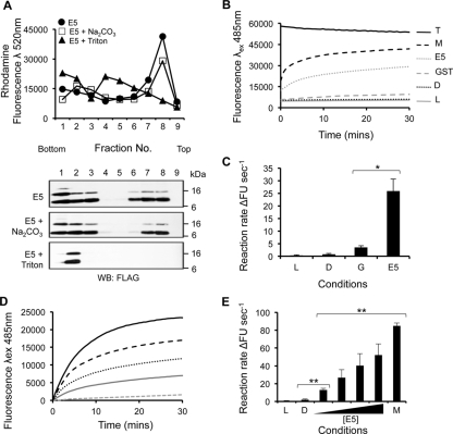
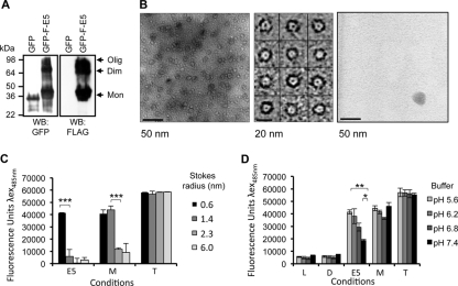
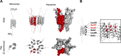
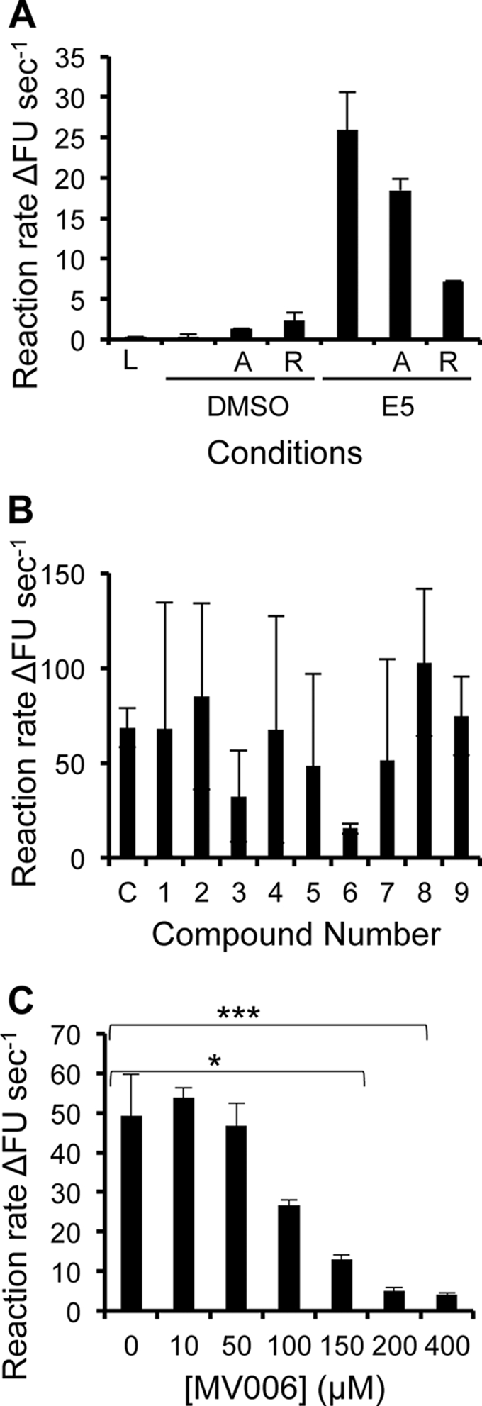
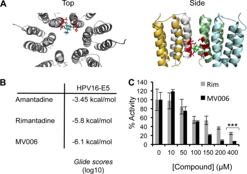
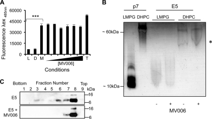
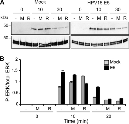
High-risk human papillomavirus type 16 (HPV16) is the primary causative agent of cervical cancer and therefore is responsible for significant morbidity and mortality worldwide. Cellular transformation is mediated directly by the expression of viral oncogenes, the least characterized of which, E5, subverts cellular proliferation and immune recognition processes. Despite a growing catalogue of E5-specific host interactions, little is understood regarding the molecular basis of its function. Here we describe a novel function for HPV16 E5 as an oligomeric channel-forming protein, placing it within the virus-encoded "viroporin" family. The development of a novel recombinant E5 expression system showed that E5 formed oligomeric assemblies of a defined luminal diameter and stoichiometry in membranous environments and that such channels mediated fluorescent dye release from liposomes. Hexameric E5 channel stoichiometry was suggested by native PAGE studies. In lieu of high-resolution structural information, established de novo molecular modeling and design methods permitted the development of the first specific small-molecule E5 inhibitor, capable of both abrogating channel activity in vitro and reducing E5-mediated effects on cell signaling pathways. The identification of channel activity should enhance the future understanding of the physiological function of E5 and could represent an important target for antiviral intervention.


Figures

Fig 1
Fig 1
Oligomerization of HPV16 E5. (A) Anti-GFP Western blot analysis of GFP and GFP-E5 immunoprecipitates from HEK293 cell lysates. A single monomeric species of GFP was observed at low levels of expression or in cells treated with the proteasome inhibitor MG132 to enhance levels of expression. Multiple bands of GFP-E5 were present, likely indicating a monomer (Mon), a dimer (Dim), and a higher-order oligomer (Olig). Note that the distribution of oligomer species did not change in cells treated with MG132. (B) Anti-FLAG Western blot analysis of FLAG precipitates from BHK cell lysates. Multiple bands of FLAG-E5 were present, including a dimer and tetramer. Note that no E5 monomer was observed in these samples. Asterisks indicate antibody heavy and light chains. Hex, hexamer. (C) FLAG immunoprecipitates (IP) from cells expressing FLAG-E5 and GFP, GFP-E5, GFP-E6, or GFP-E7. Precipitates were probed with anti-FLAG and anti-GFP antibodies to demonstrate protein interactions and equal precipitations of FLAG-E5. Analysis of lysates revealed the expression of all GFP fusion proteins. WB, Western blot. (D) FRET analysis of human cervical C33A cells coexpressing either GFP-E5 and mCherry-FLAG-E5 (i), GFP and mCherry empty vector control plasmids (ii), or GFP-E5 and mCherry empty vector control plasmids (III), pseudocolored to highlight the fluorescence intensity within each image. Images show pixel intensities following the normalization of the signal in channels, as described in Materials and Methods. (E) Hyperactivation of EGFR signaling by the E5 protein. C33A cells expressing an AP-1-responsive luciferase reporter plasmid plus untagged or GFP- or mCherry-fused E5 were mock treated or stimulated with EGF (100 ng/ml) and lysed 6 h later. Levels of luciferase were detected by luminometry and are expressed as a fold increase over mock-treated control cells. Results are the averages of data from 3 independent experiments. Error bars represent standard deviations of the means. RLU, relative light units.
Fig 2
Fig 2
Purification and analysis of recombinant HPV16 FLAG-E5. (A) Schematic of GST-FLAG-E5 designed to express the E5 protein amino-terminally fused to GST and FLAG and containing a 3C cleavage site. The FLAG tag sequence and additional residues fused to the E5 protein postcleavage are boxed. (B) SDS-PAGE and Western blot analysis of expressed E5 fusion proteins during purification using anti-FLAG and anti-GST antibodies. Lanes 1 and 7, pre-IPTG induction; lanes 2 and 8, post-IPTG induction; lanes 3 and 9, precleavage; lanes 4 and 10, postcleavage; lanes 5 and 11, solubilized pre-HPLC sample; lanes 6 and 12, post-HPLC purified FLAG-E5.
Fig 3
Fig 3
HPV16 FLAG-E5 associates with liposomes and displays dose-dependent channel activity. (A) Recombinant FLAG-E5 was assessed for membrane associations in the presence or absence of a high pH (100 mM Na2CO3 [pH 11.4]). Anti-FLAG Western blots from a fractionated, discontinuous Ficoll gradient are shown from fractions 1 to 9. The top panel shows the rhodamine fluorescence of gradient fractions with liposomes floating to the 10% Ficoll–aqueous buffer interface. Control reaction mixtures treated with the detergent Triton X-100 resulted in FLAG-E5 remaining at the bottom of the gradient. (B) Liposome assay controls. Data from a real-time analyses of the effects of liposomes only, the solvent control (DMSO), GST, FLAG-E5, melittin, and Triton X-100 on the kinetics of CF release are shown. (C) Initial rates calculated from the linear part of the real-time curve highlight the negligible impact of solvent alone (DMSO [D]) or GST (G) on CF release in comparison to FLAG-E5 (E5). Error bars represent standard deviations of the means, and the statistical significance of CF release was assessed by a one-way analysis of variance (ANOVA) comparing GST alone to FLAG-E5 (*, P < 0.05). (D and E) Increasing concentrations of recombinant FLAG-E5 ranging from 0.1 to 1 mM were incubated with CF-containing liposomes, and dye release was measured in real time (D) and by initial rates calculated from the linear part of the real-time curve (E). L, liposome only; D, DMSO solvent control; M, melittin. Error bars represent standard deviations of the means, and the statistical significance of CF release was assessed by a one-way ANOVA compared to solvent controls and E5 (**, P < 0.01).
Fig 4
Fig 4
Characterization of the HPV16 E5 channel. (A) Oligomerization of the GFP-His10-FLAG-E5 fusion protein. GFP-His10-FLAG-E5, expressed in E. coli, was analyzed by SDS-PAGE and Western blotting with antibodies against GFP or FLAG. In addition to monomeric GFP-His10-FLAG-E5, higher-molecular-weight forms were evident, representing oligomers of E5 formed in the presence of bacterial lipids. In comparison, the GFP-His10 fusion partner was present as a monomer. (B) Transmission electron microscopy of the GFP-His10-FLAG-E5 channel. This typical view from a raw image of GFP-His10-FLAG-E5 oligomers negatively stained with phosphotungstic acid (PTA) at a ×52,000 magnification (left) demonstrates the presence of “ringlike” structures only in the presence of E5. A selected set of individual GFP-His10-FLAG-E5 oligomers (middle) details the ringlike arrangement of the protein. Shown is a view of a raw TEM image of GFP-His10 negatively stained with PTA. (C) FLAG-E5 forms a channel with a restrictive luminal diameter. Liposomes containing CF or FITC-dextrans (FDs) of various molecular masses (0.38 to 70 kDa) and Stokes' radii (0.6 to 6.0 nm) were incubated with FLAG-E5 (E5) (1 μM) and melittin (M) (1 μM). Liposome-free supernatants were assessed by using fluorimetry at an λex of 485 nm and an λem 520 nm. Liposome-only and solvent controls corresponded to baseline fluorescence, and Triton X-100 controls (T) established maximum fluorescence. Baseline controls were subtracted from the fluorescence readings generated by the E5- and melittin-mediated fluorophore release. Melittin allowed the efficient release of CF and FD-4, whereas E5 would permit only the release of CF. Error bars represent standard deviations of the means. The statistical significance of E5-mediated CF release compared to 4-kDa dextran release and melittin-mediated 4-kDa and 10-kDa dextran release was assessed by a one-way ANOVA (***, P < 0.001). (D) FLAG-E5-mediated CF release from liposomes is enhanced at acidic pH. FLAG-E5 and melittin were added to CF liposomes resuspended in citrate-phosphate buffers of various pHs (5.6 to 7.4). Liposome-free supernatants were assessed by fluorimetry, and the pH of each supernatant was readjusted to pH 7.4 by the addition of buffering amounts of 1 M Tris-HCl (pH 8.0) (amounts determined by the fluorescence recovery of Triton X-100 controls). Triton X-100-lysed controls were used to determine maximum fluorescence, and baseline fluorescence was determined with liposome-alone (L) and solvent (DMSO [D]) controls. Error bars represent standard deviations of the means. The statistical significances of differences in E5-mediated CF release between pH 6.8 to 7.4 and pH 5.6 to 7.4 were assessed by a one-way ANOVA (**, P < 0.01; *, P < 0.05).
Fig 5
Fig 5
Molecular modeling of full-length HPV16 E5 as a monomer and hexamer. Models of the E5 channel complex were generated by using Maestro as described in Materials and Methods. (A) E5 monomers were modeled with free-energy minimization using the Maestro program and then manually docked into a symmetrical hexameric complex. The top panel shows a side projection (two monomers showing), and the bottom panel shows a top-down projection revealing the channel lumen. (B) Residues from TM2 that are predicted to line the lumen of the channel are highlighted.
Fig 6
Fig 6
HPV16 FLAG-E5 exhibits mixed sensitivity to adamantane compounds in vitro but can be inhibited, in a dose-dependent manner, by a novel inhibitor. (A) The sensitivity of FLAG-E5 to adamantane drugs was assessed in vitro (1 μM FLAG-E5 displayed resistance to amantadine [denoted A] but was sensitive to rimantadine [denoted R]). DMSO and FLAG-E5 in the absence of compound (denoted C) showed background and maximum activities, respectively. Error bars represent standard deviations of the means, and the statistical significance of CF release was assessed by a one-way ANOVA compared to solvent controls and FLAG-E5 (*, P < 0.05; **, P < 0.01). (B) Effects of candidate E5 inhibitors on FLAG-E5-mediated CF release. The ability of FLAG-E5 to cause the release of CF in the presence of candidate inhibitory compounds (400 μM) was assessed in real time by fluorimetry and is represented as an initial rate. Error bars represent standard deviations of the means. (C) Dose-dependent inhibition of FLAG-E5-mediated CF release by the compound MV006, as depicted by the initial rate. Error bars represent standard deviations of the means, and the statistical significance of CF release was assessed by a one-way ANOVA compared to the control of FLAG-E5 in the absence of compound (*, P < 0.05; ***, P < 0.001).
Fig 7
Fig 7
Molecular modeling of HPV16 E5 inhibitor compounds. (A) Molecular model of E5 indicating the position of the rimantadine-binding site. The binding pocket is aligned with lipophilic residues on adjacent trans-membrane domains (Ile44, Leu48, Leu45, Leu23, Leu47, Ile51, Leu48, and Ser41). The left panel shows the top view for rimantadine bound to E5. The right panel indicates a side view of rimantadine bound to E5. For simplicity, only one rimantadine molecule is shown. (B) Predicted ligand binding scores for amantadine, rimantadine, and MV006 against a hexameric E5 molecular model. (C) Comparison of the dose-dependent inhibition of FLAG-E5-mediated CF release by MV006 (black bars) and rimantadine (gray bars) highlighting the differences between the two inhibitors (***, P < 0.001).
Fig 8
Fig 8
Effects of a novel E5 inhibitor on melittin-mediated CF release, FLAG-E5 oligomerization, and membrane insertion. (A) MV006 does not prevent the melittin-induced release of CF from liposomes, as depicted by an endpoint analysis. Error bars represent standard deviations of the means, and the statistical significance of CF release was assessed by a one-way ANOVA compared to the melittin (M) control in the absence of compound (***, P < 0.001). Initial rates for liposomes alone (L), the DMSO solvent control (D), and Triton X-100 (T) are also shown. (B) Oligomerization of the FLAG-E5 protein. Mild detergents were tested for their abilities to induce the oligomerization of FLAG-p7 and FLAG-E5 using native PAGE. DHPC induced the oligomerization of both FLAG-p7 and FLAG-E5, whereas LMPG did not. The addition of MV006 (4 mM) did not impair FLAG-E5 oligomerization. The a asterisk indicates the detergent front. (C) Membrane association of FLAG-E5 in the presence of MV006. Anti-FLAG Western blots from fractionated discontinuous Ficoll gradients are shown from fractions 1 to 9. The presence of MV006 (4 mM) had no inhibitory effect on membrane insertion.
Fig 9
Fig 9
Viroporin inhibitors decrease ERK phosphorylation in E5-expressing cells. (A) HaCaT cells stably expressing HPV16 E5 or an empty plasmid were preincubated with MV006 (100 mM), rimantadine (100 mM), or the DMSO carrier for 1 h prior to stimulation with recombinant EGF (100 ng/ml). Cells were lysed, and equal amounts of protein were analyzed by SDS-PAGE. The levels of phosphorylated ERK (P-ERK) were determined by Western blotting. (B) Quantitative analysis of data from panel A using ImageJ software analysis to compare levels of phosphorylated ERK to total levels of ERK.

References

    1. Ashrafi GH, Haghshenas MR, Marchetti B, O'Brien PM, Campo MS. 2005. E5 protein of human papillomavirus type 16 selectively downregulates surface HLA class I. Int. J. Cancer 113:276–283 - PubMed
    1. Chang JL, et al. 2001. The expression of HPV-16 E5 protein in squamous neoplastic changes in the uterine cervix. J. Biomed. Sci. 8:206–213 - PubMed
    1. Clarke D, et al. 2006. Evidence for the formation of a heptameric ion channel complex by the hepatitis C virus p7 protein in vitro. J. Biol. Chem. 281:37057–37068 - PMC - PubMed
    1. Clifford GM, Smith JS, Plummer M, Munoz N, Franceschi S. 2003. Human papillomavirus types in invasive cervical cancer worldwide: a meta-analysis. Br. J. Cancer 88:63–73 - PMC - PubMed
    1. Disbrow GL, Hanover JA, Schlegel R. 2005. Endoplasmic reticulum-localized human papillomavirus type 16 E5 protein alters endosomal pH but not trans-Golgi pH. J. Virol. 79:5839–5846 - PMC - PubMed

Publication types

MeSH terms

LinkOut - more resources