

doi: 10.1128/JCM.06460-11.
Epub 2012 Feb 22.
Rapid and sensitive detection of Mycobacterium ulcerans by use of a loop-mediated isothermal amplification test
Affiliations
- PMID: 22357495
- PMCID: PMC3347114
- DOI: 10.1128/JCM.06460-11
Item in Clipboard
Rapid and sensitive detection of Mycobacterium ulcerans by use of a loop-mediated isothermal amplification test
J Clin Microbiol.
2012 May.
Abstract
This work reports the design and evaluation of a rapid loop-mediated isothermal amplification test for detecting Mycobacterium ulcerans DNA based on the multicopy insertion sequence IS2404. The test is robust and specific with a detection limit equivalent to 20 copies of the target sequence (0.01 to 0.1 genome). The test has potential for the diagnosis of Buruli ulcer under field conditions.
Figures
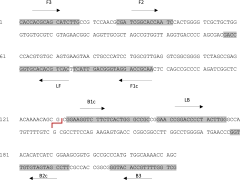
The partial nucleotide sequence of a multicopy insertion sequence, IS2404, showing the most sensitive LAMP primer set. The restriction enzyme HhaI site is shown as a red line. The primers are as follows: F3, forward; B3, backward; LF, loop forward; LB, loop backward; FIP, forward inner (F1c and F2); BIP, backward inner (B2c and B1c).

(A) The visual appearance of the BU LAMP amplification product after addition of a 1:10 dilution of SYBR green I dye. The dye fluoresces strongly when bound to the double-stranded DNA, and the resulting DNA-dye complex gives a green color, while fluorescence is minimal when the dye is free in the solution and gives an orange/brown color. (B) The amplification curves obtained using the ESE-Quant tube scanner. Results can be read using the LCD panel as either positive or negative and/or in real time using a computer with the appropriate software (9). The graph reports the fluorescence in millivolts (mV) on the y axis and time in minutes on the x axis. Sample 1, M. ulcerans-spiked soil; 2, supernatant from positive specimen; 3, DNA from needle aspirate; 4, M. marinum; control (C), M. ulcerans DNA (Agy99); negative control (NC), water.
Similar articles
-
Detection of Mycobacterium ulcerans with IS2404 loop-mediated isothermal amplification and a fluorescent reporter probe.Appl Environ Microbiol. 2025 May 21;91(5):e0027025. doi: 10.1128/aem.00270-25. Epub 2025 Apr 16. Appl Environ Microbiol. 2025. PMID: 40237451 Free PMC article.
-
Detection of Mycobacterium ulcerans by the loop mediated isothermal amplification method.PLoS Negl Trop Dis. 2012;6(4):e1590. doi: 10.1371/journal.pntd.0001590. Epub 2012 Apr 3. PLoS Negl Trop Dis. 2012. PMID: 22509415 Free PMC article.
-
Evaluation of an electricity-independent method for IS2404 Loop-mediated isothermal amplification (LAMP) diagnosis of Buruli ulcer in resource-limited settings.PLoS Negl Trop Dis. 2024 Aug 14;18(8):e0012338. doi: 10.1371/journal.pntd.0012338. eCollection 2024 Aug. PLoS Negl Trop Dis. 2024. PMID: 39141676 Free PMC article.
-
Laboratory diagnosis of Buruli ulcer disease.Future Microbiol. 2010 Mar;5(3):363-70. doi: 10.2217/fmb.10.3. Future Microbiol. 2010. PMID: 20210548 Review.
-
Use of loop-mediated isothermal amplification of DNA for the rapid detection of Mycobacterium tuberculosis in clinical specimens.Eur J Clin Microbiol Infect Dis. 2011 Aug;30(8):937-42. doi: 10.1007/s10096-011-1195-0. Epub 2011 Feb 18. Eur J Clin Microbiol Infect Dis. 2011. PMID: 21331481 Review.
Cited by
-
Real-time fluorescence loop mediated isothermal amplification for the detection of Acinetobacter baumannii.PLoS One. 2013 Jul 2;8(7):e66406. doi: 10.1371/journal.pone.0066406. Print 2013. PLoS One. 2013. PMID: 23843955 Free PMC article.
-
Stem loop-mediated isothermal amplification test: comparative analysis with classical LAMP and PCR in detection of Entamoeba histolytica in Kenya.BMC Res Notes. 2017 Mar 31;10(1):142. doi: 10.1186/s13104-017-2466-3. BMC Res Notes. 2017. PMID: 28359328 Free PMC article.
-
A Systematic Review on Suitability of Molecular Techniques for Diagnosis and Research into Infectious Diseases of Concern in Resource-Limited Settings.Curr Issues Mol Biol. 2022 Sep 21;44(10):4367-4385. doi: 10.3390/cimb44100300. Curr Issues Mol Biol. 2022. PMID: 36286015 Free PMC article. Review.
-
Genotyping Tools for Mycobacterium ulcerans-Drawbacks and Future Prospects.Mycobact Dis. 2014 May 5;4(2):1000149. doi: 10.4172/2161-1068.1000149. Mycobact Dis. 2014. PMID: 24900947 Free PMC article.
-
Establishment of reverse transcription loop-mediated isothermal amplification for rapid detection and differentiation of canine distemper virus infected and vaccinated animals.Infect Genet Evol. 2015 Jun;32:102-6. doi: 10.1016/j.meegid.2015.03.002. Epub 2015 Mar 10. Infect Genet Evol. 2015. PMID: 25769803 Free PMC article.
References
Publication types
MeSH terms
Substances
LinkOut - more resources
Full Text Sources
