. 2012 Feb 22;4(122):122ra22.
doi: 10.1126/scitranslmed.3003441.

MicroRNA-21 blocks abdominal aortic aneurysm development and nicotine-augmented expansion

Affiliations

MicroRNA-21 blocks abdominal aortic aneurysm development and nicotine-augmented expansion

Lars Maegdefessel et al. Sci Transl Med. .

Abstract

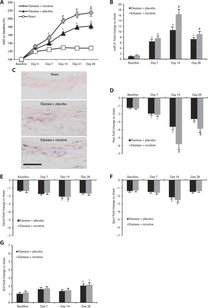
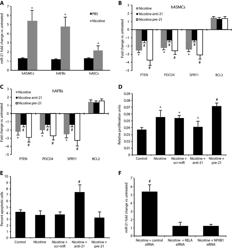
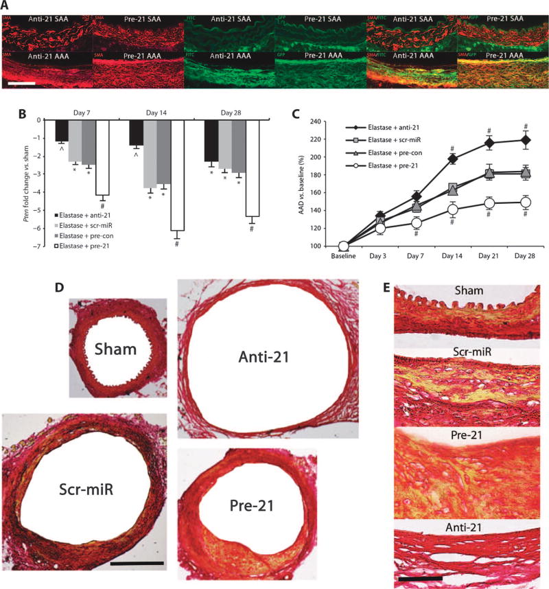
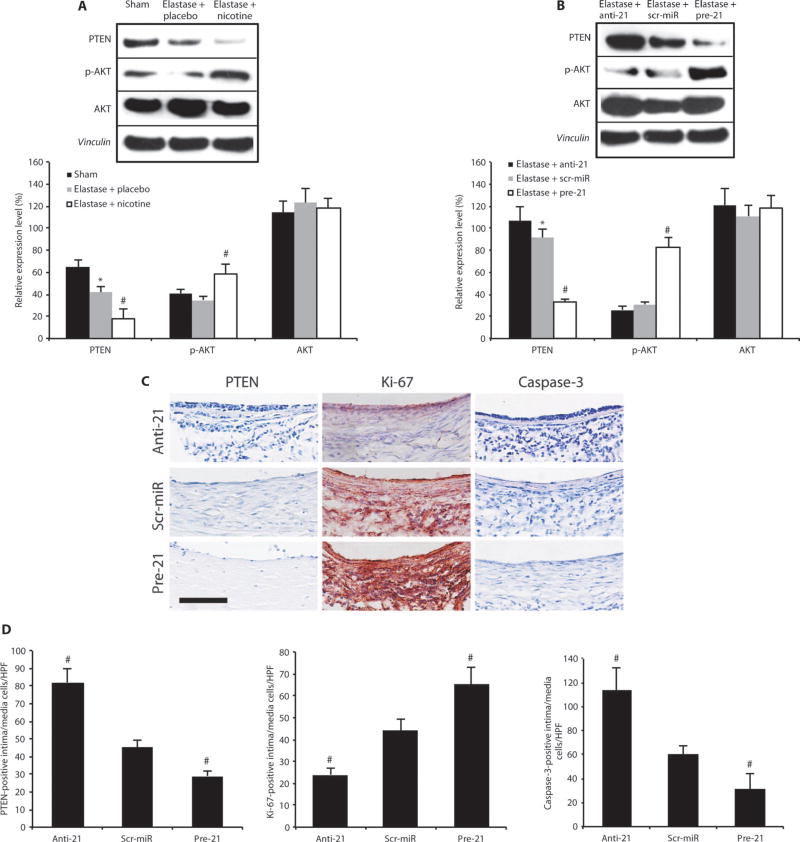
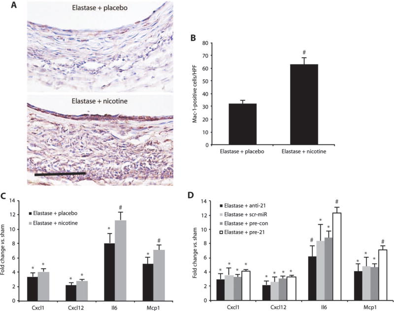
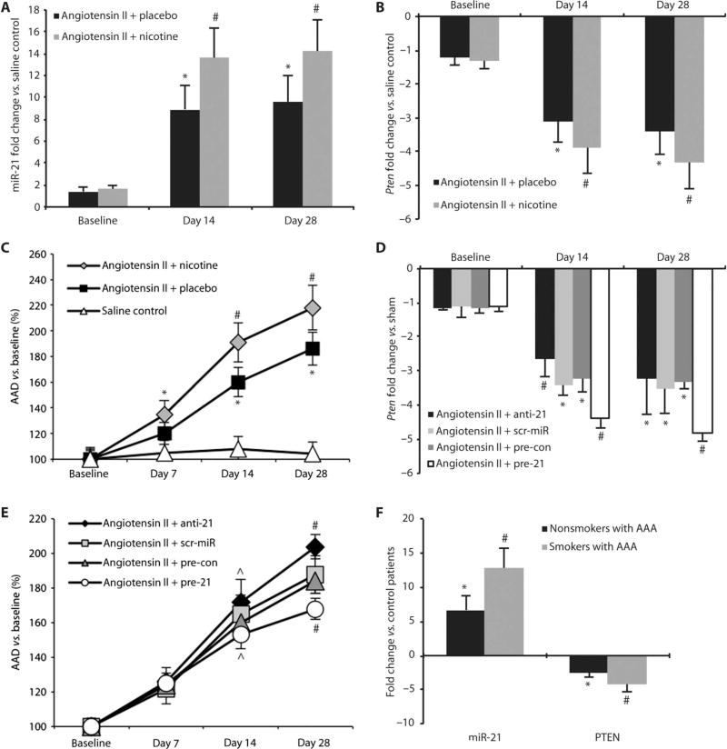
Identification and treatment of abdominal aortic aneurysm (AAA) remains among the most prominent challenges in vascular medicine. MicroRNAs are crucial regulators of cardiovascular pathology and represent possible targets for the inhibition of AAA expansion. We identified microRNA-21 (miR-21) as a key modulator of proliferation and apoptosis of vascular wall smooth muscle cells during development of AAA in two established murine models. In both models (AAA induced by porcine pancreatic elastase or infusion of angiotensin II), miR-21 expression increased as AAA developed. Lentiviral overexpression of miR-21 induced cell proliferation and decreased apoptosis in the aortic wall, with protective effects on aneurysm expansion. miR-21 overexpression substantially decreased expression of the phosphatase and tensin homolog (PTEN) protein, leading to increased phosphorylation and activation of AKT, a component of a pro-proliferative and antiapoptotic pathway. Systemic injection of a locked nucleic acid-modified antagomir targeting miR-21 diminished the pro-proliferative impact of down-regulated PTEN, leading to a marked increase in the size of AAA. Similar results were seen in mice with AAA augmented by nicotine and in human aortic tissue samples from patients undergoing surgical repair of AAA (with more pronounced effects observed in smokers). Modulation of miR-21 expression shows potential as a new therapeutic option to limit AAA expansion and vascular disease progression.


Conflict of interest statement

Competing interests: L.M., J.M.S., and P.S.T. have applied for a patent relating to miR-21 and protection from AAA development. The other authors declare that they have no competing interests.

Figures

Fig. 1
Fig. 1
miR-21 and target gene expression in AAAs induced by elastase infusion. (A) AAD (versus baseline as a percentage) in elastase-infused mice supplemented with placebo and nicotine compared to saline-infused control mice (sham). (B) miR-21 expression in mice infused with elastase supplemented with placebo or nicotine compared to sham animals. (C) ISH staining formiR-21 (purple chromagen) in mice infused with elastase and supplemented with placebo, elastase-infused mice supplemented with nicotine, or saline-infused (sham) animals 14 days after AAA induction (labels on luminal side; scale bar, 50 µm). (D to G) mRNA expression for Pten (D), Pdcd4 (E), Spry1 (F), and Bcl2 (G) genes in mice infused with elastase and supplemented with either placebo or nicotine compared to sham animals. n = 5 to 8 for each treatment group and time point. Data are means ± SEM. *P < 0.05 versus sham; #P < 0.05 versus elastase + placebo and sham. Level of significance was determined using one-way ANOVA with Bonferroni’s post test. Sequential measurements (AADs at consecutive time points) were analyzed by one-way repeated-measures ANOVA.
Fig. 2
Fig. 2
Effects of nicotine and miR-21 modulation in vitro. (A) miR-21 expression in nicotine (10 nM)– and PBS-treated (control) cultured hASMCs, hAFBs, and hAECs. (B and C) Expression of miR-21 target genes in nicotine-treated hASMCs (B) or hAFBs (C) transfected with miRNAs that block (anti-21) or boost (pre-21) miR-21 expression. (D) Proliferation of hASMCs after nicotine and pre-21 treatment was measured using the MTT assay. (E) Apoptosis of hASMCs after anti-21 treatment was measured by detecting the percent of annexin V–positive cells. (F) Knockdown of NF-κB subunits (RELA and NFKB1) with siRNA in nicotine-treated hASMCs. Data are means ± SEM. *P < 0.05 versus untreated control; #P < 0.05 for anti-21 andpre-21 (+nicotine) versus scr-miR and saline control (or nicotine + control siRNA versus nicotine + RELA/NFKB1 siRNA and versus untreated); ^P < 0.05 versus pre-21, scr-miR, and nicotine alone. Level of significance was determined using one-way ANOVA with Bonferroni’s post test.
Fig. 3
Fig. 3
Effects of anti-21 and pre-21 in vivo. (A) Double immunofluorescence images for SMA (red) and GFP/FITC label (green) in the non-aneurysmal part of the suprarenal abdominal aorta (SAA) and in the infrarenal site of injury (AAA) in AAAs from mice treated with anti-21 or pre-21 (as single-color and merged images; labels on luminal side; scale bar, 50 µm). (B) Pten expression in elastase-induced AAA after treatment with anti-21, pre-21, scr-miR, or empty vector control (pre-con) compared to saline-infused mice (sham). (C) AAD (versus baseline as a percentage) in anti-21– and pre-21–transduced mice compared to scr-miR and empty vector control (pre-con) in the elastase infusion model of AAA. (D and E) Representative low-power [(D) scale bar, 400 µm] and high-power [(E) scale bar, 50 µm] images of aortic cross sections stained with Picrosirius Red to illustrate differences in vascular wall structure (yellow/orange, muscle; red, collagen) in the aortic wall in anti-21– and pre-21–transduced mice compared to saline-infused controls (sham) and scr-miR in the elastase infusion model of AAA 28 days after induction. Scale bar, 50 µm. n = 4 to 8 mice for each time point and group. Data are means ± SEM. *P < 0.05 versus sham; #P < 0.05 versus scr-miR/pre-con and sham; ^P < 0.05 versus sham and pre-21. Level of significance was determined using one-way ANOVA with Bonferroni’s post test. Sequential measurements (AADs at consecutive time points) were analyzed by one-way repeated-measures ANOVA.
Fig. 4
Fig. 4
PTEN and p-AKT regulate proliferation and apoptosis in AAAs. (A) Representative immunoblots and expression levels for PTEN, p-AKT, and AKT relative to vinculin (loading control) in AAAs in elastase-infused mice (14 days after AAA induction) supplemented with either placebo or nicotine compared to saline-infused controls (sham; n = 3 to 4 mice per group). (B) Representative immunoblots and expression levels for PTEN, p-AKT, and AKT relative to vinculin (loading control) in AAAs in elastase-infusedmice (14 days after induction) transduced with anti-21 or pre-21 compared with scr-miR (n = 3 to 4 mice per group). (C) Representative immunohistochemical images demonstrating effects of treatment with anti-21, pre-21, or scr-miR 14 days after AAA induction with elastase on PTEN expression (blue), cell proliferation (red positive for Ki-67), and apoptosis (purple-blue positive for caspase-3). Scale bar, 50 µm. (D) Quantification of PTEN-positive, Ki-67–positive, and caspase-3–positive cells in the intimal and medial region of AAAs. n=4 high-power fields (HPF) of 3 different aortas per group (12 total per group) 14 days after AAA induction. Data are means ± SEM. *P < 0.05 versus saline-infused controls (sham); #P < 0.05 versus scr-miR and versus sham (or versus elastase + placebo and versus sham). Level of significance was determined using one-way ANOVA with Bonferroni’s post test.
Fig. 5
Fig. 5
Nicotine triggers inflammation. (A) Mac-1–positive cells (red) are increased in mice supplemented with nicotine compared to placebo in the elastase-induced mouse model of AAAs. Scale bar, 50 µm. (B) Mac-1–positive cells from n = 4 high-power fields (HPF) from 3 different mice per group (12 total per group). (C) Gene expression of Cxcl1, Cxcl12, Il6, and Mcp1 in mice infused with elastase and supplemented with either nicotine or placebo compared with saline-infused control mice (sham). (D) Gene expression of Cxcl1, Cxcl12, Il6, and Mcp1 in mice transduced with anti-21 or pre-21, scr-miR, or empty vector control (pre-con) compared to sham 14 days after induction of AAA by elastase infusion. n = 3 to 7 per treatment group. Data are means ± SEM. *P < 0.05 versus sham; #P < 0.05 versus scr-miR, empty vector control (pre-con), and sham (or versus elastase + placebo and versus sham). Level of significance was determined using one-way ANOVA with Bonferroni’s post test, or Student’s t test for parametric measures (B).
Fig. 6
Fig. 6
miR-21 and PTEN expression in mouse and human AAAs. (A) miR-21 expression in nicotine-treated and placebo-implanted mice infused with angiotensin II to induce AAAs compared to saline-infused control mice. (B) Pten expression in nicotine-treated and placebo-implanted mice infused with angiotensin II to induce AAAs compared to saline-infused control mice. (C) Expansion (as a percentage of baseline) of the AAD for nicotine-treated (n = 35) and placebo-implanted mice (n = 28) infused with angiotensin II to induce AAAs compared to saline-infused control mice (n = 18). (D) Pten expression in anti-21– and pre-21–transduced mice, mice treated with scr-miR, or empty vector control (pre-con) compared to saline-infused control mice in the angiotensin II model of AAA. (E) AAD (as a percentage of baseline) in anti-21– and pre-21–transduced mice, as well as mice treated with scr-miR or empty vector control (pre-con) in the angiotensin II model of AAA. (F) Up-regulation of miR-21 and down-regulation of PTEN in human AAA tissue (n = 5 nonsmokers, n = 8 smokers) undergoing surgical repair compared to aortic samples from control patients without AAA (n = 5). Data are means ± SEM. *P < 0.05 versus saline controls (or control patients); #P < 0.05 versus placebo with angiotensin II and saline controls, scr-miR or pre-con, or nonsmokers and control patients; ^P < 0.05 versus angiotensin II with anti-21 or pre-21. Level of significance was determined using one-way ANOVA with Bonferroni’s post test. Sequential measurements (AADs at consecutive time points) were analyzed by one-way repeated-measures ANOVA.

Similar articles

Cited by

References

    1. Fleming C, Whitlock EP, Beil TL, Lederle FA. Screening for abdominal aortic aneurysm: A best-evidence systematic review for the U.S. Preventive Services Task Force. Ann. Intern. Med. 2005;142:203–211. - PubMed
    1. Creager MA, Jones DW, Easton JD, Halperin JL, Hirsch AT, Matsumoto AH, O’Gara PT, Safian RD, Schwartz GL, Spittell JA American Heart Association, Atherosclerotic Vascular Disease Conference: Writing Group V. Medical decision making and therapy. Circulation. 2004;109:2634–2642. - PubMed
    1. Powell JT, Worrell P, MacSweeney ST, Franks PJ, Greenhalgh RM. Smoking as a risk factor for abdominal aortic aneurysm. Ann. N. Y. Acad. Sci. 1996;800:246–248. - PubMed
    1. Weintraub NL. Understanding abdominal aortic aneurysm. N Engl. J. Med. 2009;361:1114–1116. - PMC - PubMed
    1. Lederle FA, Nelson DB, Joseph AM. Smokers’ relative risk for aortic aneurysm compared with other smoking-related diseases: A systematic review. J Vasc. Surg. 2003;38:329–334. - PubMed

Publication types

MeSH terms

LinkOut - more resources