Review
. 2012 May 31;86(5):162, 1-14.
doi: 10.1095/biolreprod.111.095463. Print 2012 May.

Male germline control of transposable elements

Affiliations
Review

Male germline control of transposable elements

Jianqiang Bao et al. Biol Reprod. .

Abstract

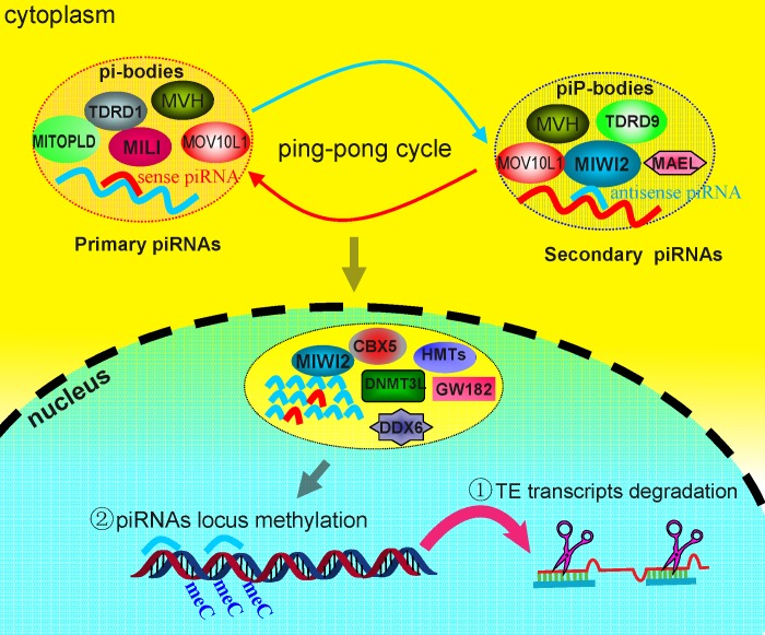
Repetitive sequences, especially transposon-derived interspersed repetitive elements, account for a large fraction of the genome in most eukaryotes. Despite the repetitive nature, these transposable elements display quantitative and qualitative differences even among species of the same lineage. Although transposable elements contribute greatly as a driving force to the biological diversity during evolution, they can induce embryonic lethality and genetic disorders as a result of insertional mutagenesis and genomic rearrangement. Temporary relaxation of the epigenetic control of retrotransposons during early germline development opens a risky window that can allow retrotransposons to escape from host constraints and to propagate abundantly in the host genome. Because germline mutations caused by retrotransposon activation are heritable and thus can be deleterious to the offspring, an adaptive strategy has evolved in host cells, especially in the germline. In this review, we will attempt to summarize general defense mechanisms deployed by the eukaryotic genome, with an emphasis on pathways utilized by the male germline to confer retrotransposon silencing.


Figures

FIG. 1.
FIG. 1.
Schematic depiction of different classes of transposable elements (TEs) in the mammalian genome. Transposable elements can be divided overall into DNA transposons (class II; A) and retrotransposons (class I; BE), according to their sequence features and mode of transposition. Most mammalian genomes are occupied by the retrotransposons, which can be further subcategorized into long terminal repeat (LTR) retrotransposons with LTRs at both ends (B) and non-LTR retrotransposons, including LINEs (long interspersed nucleotide elements) (C), SINEs (short interspersed nucleotide element) (D), and SVAs (E). LTR retrotransposons, also known as endogenous retrovirus (ERV), can encode two proteins, POL and GAG, but lack the coding sequence of ENV protein that is critical for the full life cycle of the retrovirus. LINE1, with the typical full-length structure being shown in C, is capable of translating two ORFs: ORF1, encoding a RNA-binding protein, and ORF2, encoding two enzymes—reverse transcriptase and endonuclease—that are essential for autonomous movement of LINEs. The insidious promoter residing in the 5′-UTR drives the transcription of non-LTR LINE by DNA polymerase II, whereas SINEs, of which a typical representative is Alu (D), comprises ∼18% of the human genome and are short DNA sequence that cannot encode any functional reverse transcriptase protein and thus rely on other mobile elements for transposition. SVAs (E) contain two typical sequence motifs, namely, Alu-like and SINE-R, which are separated by a variable number of tandem repeats (VNTR) locus. To date, active TEs currently known include LINEs, SINEs and SVAs. LINEs can transpose through autonomous mobilization, whereas both SINEs and SVAs depend on LINE1 activity for nonautonomous transposition.
FIG. 2.
FIG. 2.
Schematic representation of two genomic reprograming events during murine development. Genomic methylation levels (vertical axis) during preimplantation and fetal germ cell development (horizontal axis) in mice are shown. Blue and red lines represent paternal and maternal genomes, respectively. Dashed lines represent the progression of demethylation. Developmental time points are indicated on the horizontal axis. A) Fertilization: demethylation of the paternal genome initiates immediately after the formation of the pronucleus, while methylation of the maternal genome is unchanged. B) First cleavage: demethylation of the maternal genome commences, whereas the paternal genome demethylation is complete prior to the first cleavage. C) Blastomere stage: there is a notable passive demethylation in the maternal genome concomitant with embryo cleavages between stages B and C. D) At 3.5 dpc: the embryo is at the blastocyst stage. Both paternal and maternal genomes start the remethylation process. E) At ∼4.5 dpc: methylation levels of both paternal and maternal genomes peak in the embryo. F) At 7.5 dpc: PGCs appear in the allantois as a population of ∼40 cells and continue to proliferate and migrate toward the gonad. G) At 10.5–11.5 dpc: PGCs reach the genital ridge, and demethylation occurs in the PGC genome. H) At 13.5 dpc: PGCs cease proliferation, and sex-specific gonad differentiation is being completed. PGCs become prospermatogonia in the testis and oogonia in the ovary. Prospermatogonia continue to proliferate mitotically while oogonia proceed to meiosis. I) At 15.5 dpc: de novo methylation continues in prospermatogonia while oogonia become oocytes that are in prophase I of meiosis. J) At 17.5 dpc: methylation levels of prospermatogonia almost peak at 17.5 dpc. K) Birth: de novo remethylation of maternal genome commences at growing oocyte close to or shortly after birth in females.
FIG. 3.
FIG. 3.
Male germinal granules, including the intermitochondrial cement and the chromatoid body, are present in the cytoplasm during male germline development in the mouse. Spermatogenic cells containing the intermitochondrial cement are framed with a blue dashed line, whereas the red dashed-line frame indicates spermatogenic cells possessing the chromatoid body. Note that both the intermitochondrial cement and the chromatoid body are present in late pachytene spermatocytes.
FIG. 4.
FIG. 4.
Schematic summary of expression profiles of the PIWI-piRNAs pathway components and currently known male germ cell-associated epigenetic modifiers during male germline development. Reprogramming occurs in fetal male germ cells (primordial germ cells and prospermatogonia) between 7.5 dpc and birth. DNMT3L is responsible for de novo methylation, while DNMT3A/B serve as the major maintenance methyltransferases. PIWI family proteins associate with respective TDRD members, such as MILI-TDRD1and MIWI2-TDRD9, and localize to different subcellular compartments. Moreover, these proteins most probably complex with other cofactors, such as MOV10L1, MAEL, GASZ, and HSPA2 (HSP70-2), to fulfill their functions in piRNA production and TE suppression. The height of the thick lines represents the relative expression levels of a protein at the stages indicated. Prospg, prospermatogonia; SSC, spermatogonial stem cell; Spg, spermatogonia; L, leptotene spermatocytes; P, pachytene spermatocytes; D, diplotene spermatocytes; Spd, spermatids.
FIG. 5.
FIG. 5.
Subcellular localization of components of the PIWI-piRNA pathway and the proposed mechanism of TE suppression by this pathway. MILI associates with TDRD1 and other cofactors localized to pi-bodies, whereas MIWI2 binds TDRD9 and several other P body components localized to piP-bodies. Sense and antisense piRNAs produced from both granules can exchange through the presumptive ping-pong cycle to enhance piRNAs production. MIWI2 can enter the nucleus by recruiting other epigenetic modifiers such as DNMT3L, CBX5 (HP1a), and HMTs (histone methyltransferases). The resulting secondary antisense piRNAs can presumably function to suppress transposons via two mechanisms: 1) degradation of the retrotranscripts by recognizing complementary RNA in an RNA:RNA hybrid at the posttranscriptional level and 2) recruiting epigenetic repressors responsible for DNA methylation and heterochromatin formation via an unidentified mechanism.

Similar articles

Cited by

References

    1. Biemont C, Vieira C. Genetics: junk DNA as an evolutionary force. Nature 2006; 443: 521 524 - PubMed
    1. Kidwell MG, Lisch DR. Perspective: transposable elements, parasitic DNA, and genome evolution. Evolution 2001; 55: 1 24 - PubMed
    1. Gregory TR, Hebert PD. The modulation of DNA content: proximate causes and ultimate consequences. Genome Res 1999; 9: 317 324 - PubMed
    1. Waterston RH, Lindblad-Toh K, Birney E, Rogers J, Abril JF, Agarwal P, Agarwala R, Ainscough R, Alexandersson M, An P, Antonarakis SE, Attwood J. et al. Initial sequencing and comparative analysis of the mouse genome. Nature 2002; 420: 520 562 - PubMed
    1. Kazazian HH., Jr Mobile elements: drivers of genome evolution. Science 2004; 303: 1626 1632 - PubMed

Publication types

MeSH terms