. 2012 Apr;23(8):1593-604.
doi: 10.1091/mbc.E11-11-0900. Epub 2012 Feb 22.

Rnd1 and Rnd3 targeting to lipid raft is required for p190 RhoGAP activation

Affiliations

Rnd1 and Rnd3 targeting to lipid raft is required for p190 RhoGAP activation

Izumi Oinuma et al. Mol Biol Cell. 2012 Apr.

Abstract

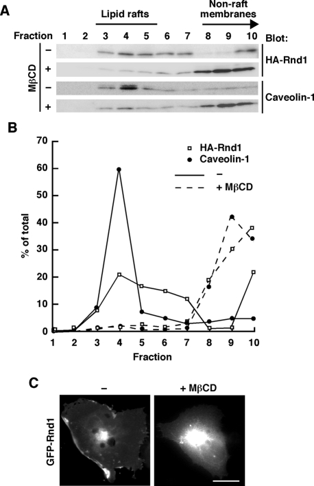
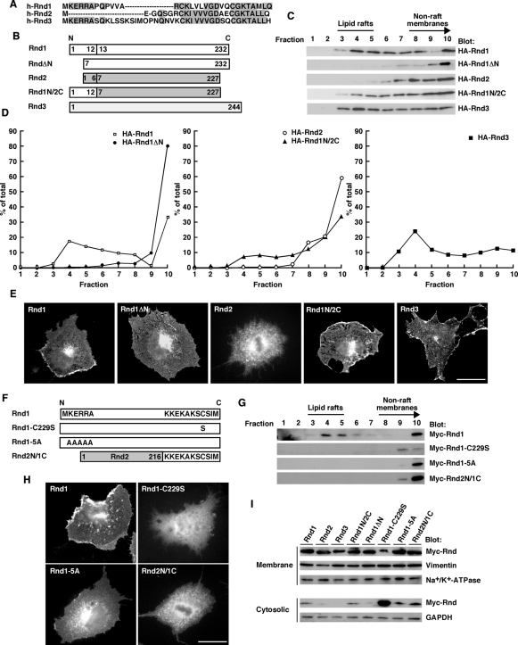
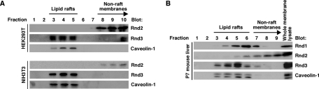
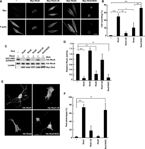
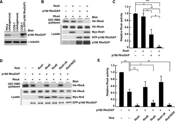
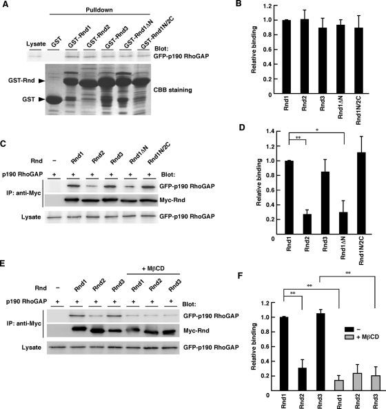
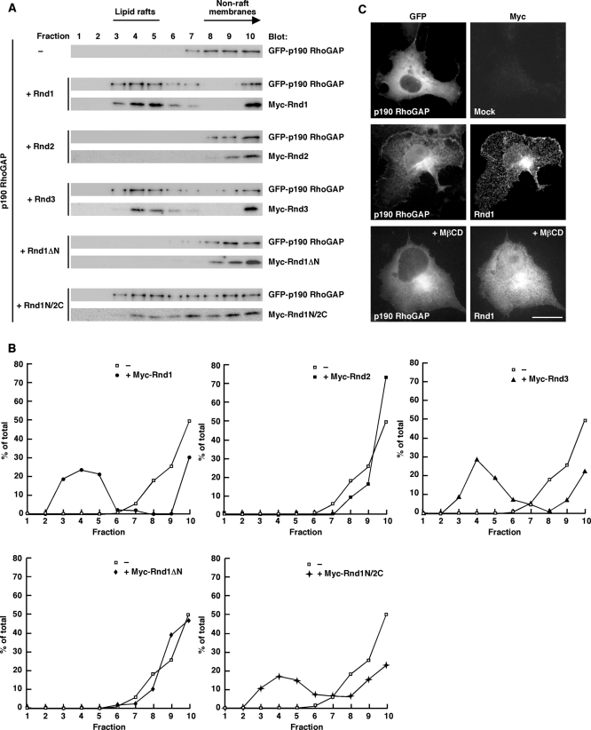
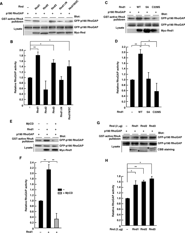
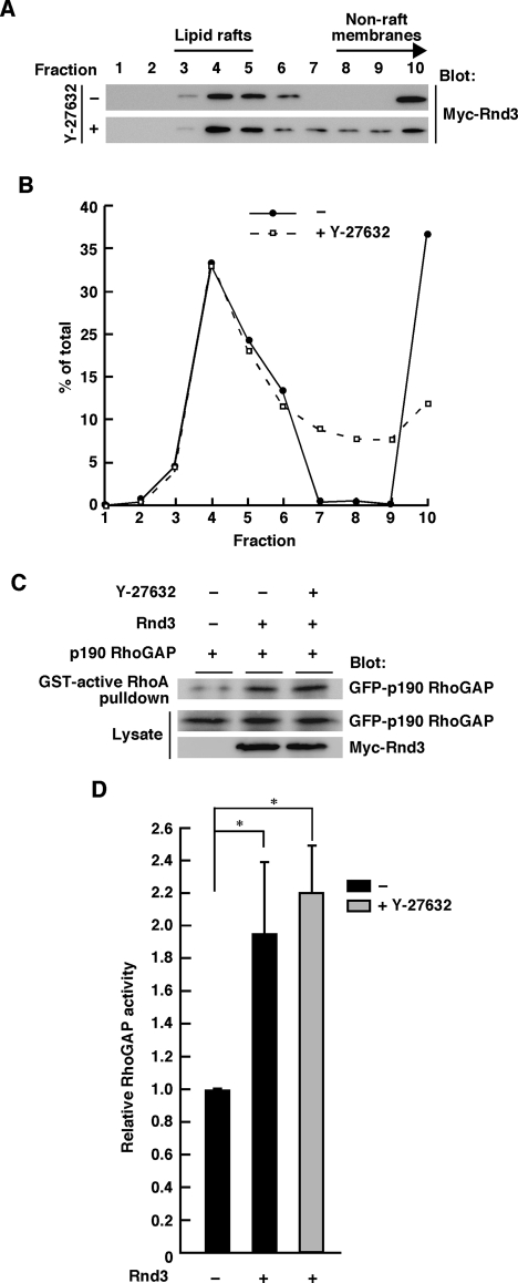
The Rnd proteins Rnd1, Rnd2, and Rnd3/RhoE are well known as key regulators of the actin cytoskeleton in various cell types, but they comprise a distinct subgroup of the Rho family in that they are GTP bound and constitutively active. Functional differences of the Rnd proteins in RhoA inhibition signaling have been reported in various cell types. Rnd1 and Rnd3 antagonize RhoA signaling by activating p190 RhoGAP, whereas Rnd2 does not. However, all the members of the Rnd family have been reported to bind directly to p190 RhoGAP and equally induce activation of p190 RhoGAP in vitro, and there is no evidence that accounts for the functional difference of the Rnd proteins in RhoA inhibition signaling. Here we report the role of the N-terminal region in signaling. Rnd1 and Rnd3, but not Rnd2, have a KERRA (Lys-Glu-Arg-Arg-Ala) sequence of amino acids in their N-terminus, which functions as the lipid raft-targeting determinant. The sequence mediates the lipid raft targeting of p190 RhoGAP correlated with its activation. Overall, our results demonstrate a novel regulatory mechanism by which differential membrane targeting governs activities of Rnd proteins to function as RhoA antagonists.


Figures

FIGURE 1:
FIGURE 1:
Lipid raft localization of Rnd1. (A) Rnd1-transfected or nontransfected COS-7 cells treated with (+) or without (–) MβCD were subjected to the sucrose density gradient fractionation assay. Each fraction (sampled and numbered from the top of the gradient) was analyzed by immunoblotting using anti-HA antibody (top) or anti–caveolin-1 antibody (bottom). (B) Densitometric analysis of the bands on the immunoblots in A. The amount of the protein in each fraction was expressed as a percentage of the total. (C) COS-7 cells were transiently transfected with GFP-Rnd1 and treated with (+) or without (–) MβCD (20 mM) before fixation. Scale bar, 20 μm.
FIGURE 2:
FIGURE 2:
The N-terminal KERRA sequence of Rnd1 mediates lipid raft targeting and plasma membrane localization. (A) The amino-terminal amino acid sequences of human Rnd1, Rnd2, and Rnd3. Identical amino acids are boxed in gray. (B) Schematic representation of the constructs used in this study. Numbers indicate amino acid positions within the sequence. (C) COS-7 cells transfected with the indicated expression plasmids were subjected to the sucrose density gradient fractionation assay. (D) Densitometric analysis of HA-Rnd bands on the immunoblots in C. The amount of the protein in each fraction was expressed as a percentage of the total. (E) COS-7 cells were transiently transfected with the Myc-tagged expression plasmids of Rnd proteins and stained with anti-Myc monoclonal antibody. Scale bar, 20 μm. (F) Schematic representation of the constructs. (G) COS-7 cells transfected with the indicated expression plasmids were subjected to the sucrose density gradient fractionation assay. (H) COS-7 cells were transiently transfected with the Myc-tagged expression plasmids of Rnd proteins and stained with anti-Myc monoclonal antibody. Scale bar, 20 μm. (I) Cellular homogenates from COS-7 cells transiently transfected with the indicated plasmids were separated into the crude membrane and the cytosolic fractions and then analyzed by immunoblotting. The fractions were immunoblotted with anti-vimentin and anti–Na+/K+-ATPase antibodies for the control membrane proteins and with anti-GAPDH antibody for the control soluble protein.
FIGURE 3:
FIGURE 3:
Rnd1 and Rnd3, but not Rnd2, reside in lipid rafts in vivo. (A) HEK293T cells (top) and NIH3T3 cells (bottom) were subjected to the sucrose density gradient fractionation assay. Ten fractions were sampled and numbered from the top of the gradient, and each fraction was analyzed by immunoblotting. (B) Liver tissue homogenate prepared from postnatal day 7 mouse was subjected to the sucrose density gradient fractionation assay. Nine fractions were sampled and numbered from the top of the gradient, and each fraction and the whole membrane lysate were analyzed by immunoblotting.
FIGURE 4:
FIGURE 4:
The N-terminal sequence of Rnd1 is required for RhoA inhibition signaling. (A) HeLa cells were transiently transfected with the indicated expression plasmids and stained with anti-Myc monoclonal antibody and with phalloidin to visualize F-actin. (B) Myc-staining–positive cells with an area <300 μm2 were scored as a percentage of the total number of transfected cells (n = 50/sample). (C) HeLa cells were transfected with the indicated expression plasmids, and the cell lysates were incubated with GST-fused, Rho-binding domain of rhotekin (GST-RBD). Bound RhoA and total cell lysates were analyzed by immunoblotting. (D) Relative RhoA activity was determined by the amount of RhoA bound to GST-RBD normalized to the amount of RhoA in cell lysates. The results are the mean ± SEM of three independent experiments. (E) Swiss 3T3 fibroblasts were microinjected with the indicated expression plasmids and stained with anti-HA monoclonal antibody. (F) Cells exhibiting the Rnd phenotype were scored as a percentage of the total number of microinjected cells. The results are the mean ± SEM of three independent experiments (n = 30/sample). Scale bars, 25 μm. ***p < 0.001, **p < 0.01, *p < 0.05 (one-way ANOVA).
FIGURE 5:
FIGURE 5:
Role of the N-terminal sequence of Rnd1 in p190 RhoGAP-mediated RhoA inhibition. (A) Whole-cell lysates from untransfected or GFP-p190 RhoGAP–transfected cells were analyzed by immunoblotting with anti–p190 RhoGAP and anti–α-tubulin antibodies. (B–E) COS-7 cells were transfected with the indicated expression plasmids, and relative RhoA activity was measured as described in the legend to Figure 4, C and D. The results are the mean ± SEM of three independent experiments. ***p < 0.001, **p < 0.01, *p < 0.05 (one-way ANOVA).
FIGURE 6:
FIGURE 6:
Lipid raft enables efficient binding of Rnd1 to p190 RhoGAP. (A) COS-7 cells were transfected with an expression vector encoding GFP-tagged p190 RhoGAP, and the cell lysate was incubated with GST-fused Rnd proteins. GST proteins used in the pulldown assay were verified by CBB staining. (C, E) Lysates from transiently transfected COS-7 cells with or without MβCD treatment (20 mM, 20 min) were immunoprecipitated with an antibody against Myc. Bound and total proteins were analyzed by immunoblotting with the indicated antibodies. (B, D, F) Relative binding was determined by the amount of bound p190 RhoGAP normalized to the amount of p190 RhoGAP in cell lysates. The results are the mean ± SEM of three independent experiments. **p < 0.01, *p < 0.05 (one-way ANOVA).
FIGURE 7:
FIGURE 7:
Rnd-dependent raft targeting of p190 RhoGAP. (A) COS-7 cells transiently transfected with the indicated plasmids were subjected to the sucrose density gradient fractionation assay. (B) Densitometric analysis of the GFP-p190 RhoGAP bands on the immunoblots in A. The amount of the GFPxp190 RhoGAP protein in each fraction was expressed as a percentage of the total. –, in the absence of Rnd1. (C) COS-7 cells were transiently transfected with GFP–p190 RhoGAP or GFP–p190 RhoGAP plus Myc-tagged Rnd1 and stained with anti-Myc monoclonal antibody. MβCD (20 mM) was treated for 20 min before fixation. Scale bar, 20 μm.
FIGURE 8:
FIGURE 8:
Rnd1 induces lipid raft–dependent p190 RhoGAP activation. (A, C, E) Functionally active p190 RhoGAP in COS-7 lysates expressing the indicated Rnd proteins was pulled down by GST-fused active RhoA. MβCD treatment (20 mM) was performed for 20 min before cell lysis. (G) COS-7 cells were transfected with an expression vector encoding GFP-tagged p190 RhoGAP, and the cell lysate was incubated with 2 μg of nonfused recombinant Rnd proteins. Rnd proteins used in the assay were verified by CBB staining (bottom, indicated by asterisks). (B, D, F, H) Relative RhoGAP activity was determined by the amount of bound p190 RhoGAP normalized to the amount of p190 RhoGAP in cell lysates. The results are the mean ± SEM of three independent experiments. **p < 0.01, *p < 0.05 (one-way ANOVA).
FIGURE 9:
FIGURE 9:
Rnd3 displays ROCK-independent lipid raft localization and activation of p190 RhoGAP. (A) Rnd3-transfected COS-7 cells treated with (+) or without (–) Y-27632 (25 μM) treatment were subjected to the sucrose density gradient fractionation assay. (B) Densitometric analysis of the bands on the immunoblots in A. The amount of the protein in each fraction was expressed as a percentage of the total. (C) Functionally active p190 RhoGAP in Rnd3-transfected COS-7 lysates with or without Y-27632 (25 μM) treatment was pulled down by GST-fused active RhoA. (D) Relative RhoGAP activity was determined by the amount of bound p190 RhoGAP normalized to the amount of p190 RhoGAP in cell lysates. The results are the mean ± SEM of three independent experiments. *p < 0.05 (one-way ANOVA).

References

    1. Barberis D, Casazza A, Sordella R, Corso S, Artigiani S, Settleman J, Comoglio PM, Tamagnone L. p190 Rho-GTPase activating protein associates with plexins and it is required for semaphoring signalling. J Cell Sci. 2005;118:4689–4700. - PubMed
    1. Brown DA, London E. Functions of lipid rafts in biological membranes. Annu Rev Cell Dev Biol. 1998;14:111–136. - PubMed
    1. Chardin P. Function and regulation of Rnd proteins. Nat Rev Mol Cell Biol. 2006;7:54–62. - PubMed
    1. del Pozo MA, Alderson NB, Kiosses WB, Chiang HH, Anderson RG, Schwartz MA. Integrins regulate Rac targeting by internalization of membrane domains. Science. 2004;303:839–842. - PubMed
    1. Foster R, Hu KQ, Lu Y, Nolan KM, Thissen J, Settleman J. Identification of a novel human Rho protein with unusual properties: GTPase deficiency and in vivo farnesylation. Mol Cell Biol. 1996;16:2689–2699. - PMC - PubMed

Publication types

LinkOut - more resources