. 2012 Apr;23(8):1475-85.
doi: 10.1091/mbc.E11-11-0957. Epub 2012 Feb 22.

Phosphorylation of threonine 1736 in the C-terminal tail of integrin β4 contributes to hemidesmosome disassembly

Affiliations

Phosphorylation of threonine 1736 in the C-terminal tail of integrin β4 contributes to hemidesmosome disassembly

Evelyne Frijns et al. Mol Biol Cell. 2012 Apr.

Abstract

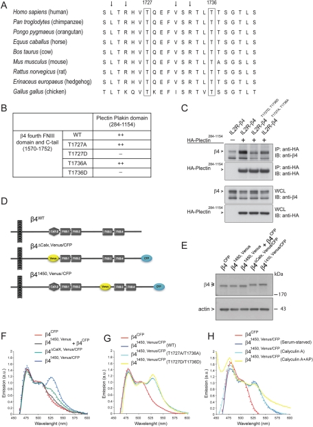
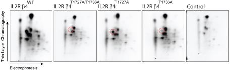
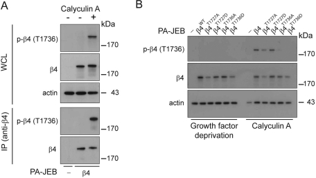
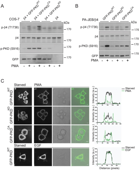
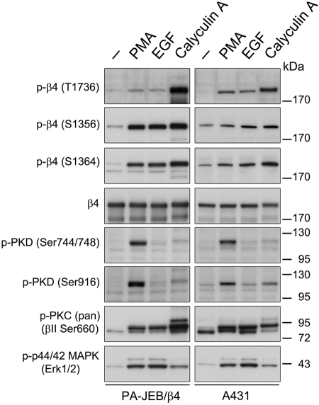
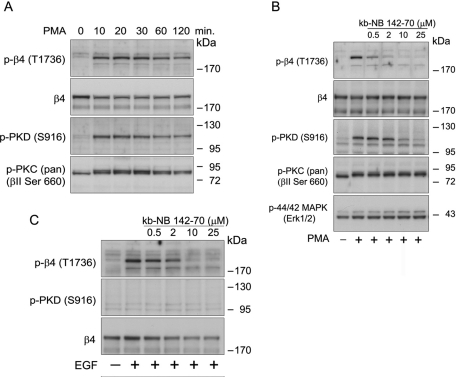
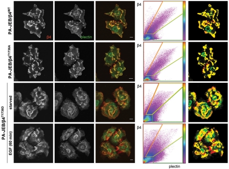
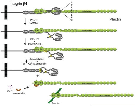
During wound healing, hemidesmome disassembly enables keratinocyte migration and proliferation. Hemidesmosome dynamics are altered downstream of epidermal growth factor (EGF) receptor activation, following the phosphorylation of integrin β4 residues S1356 and S1364, which reduces the interaction with plectin; however, this event is insufficient to drive complete hemidesmome disassembly. In the studies reported here, we used a fluorescence resonance energy transfer-based assay to demonstrate that the connecting segment and carboxy-terminal tail of the β4 cytoplasmic domain interact, which facilitates the formation of a binding platform for plectin. In addition, analysis of a β4 mutant containing a phosphomimicking aspartic acid residue at T1736 in the C-tail suggests that phosphorylation of this residue regulates the interaction with the plectin plakin domain. The aspartic acid mutation of β4 T1736 impaired hemidesmosome formation in junctional epidermolysis associated with pyloric atresia/β4 keratinocytes. Furthermore, we show that T1736 is phosphorylated downstream of protein kinase C and EGF receptor activation and is a substrate for protein kinase D1 in vitro and in cells, which requires its translocation to the plasma membrane and subsequent activation. In conclusion, we identify T1736 as a novel phosphorylation site that contributes to the regulation of hemidesmome disassembly, a dynamically regulated process involving the concerted phosphorylation of multiple β4 residues.


Figures

FIGURE 1:
FIGURE 1:
Mimicking phosphorylation of β4 on T1727 or T1736 prevents binding of the C-tail of β4 to the plakin domain of plectin. (A) Amino acid sequence (1721–1742) alignment of β4 from different species using ClustalW. Besides the residues T1727 and T1736, the leucine, valine, and arginines (arrows) are highly conserved. (B) Substitution of T1727 or T1736 on β4 by a phosphomimicking aspartic acid residue resulted in loss of binding of the fourth FNIII domain and C-tail of β4 to the plectin plakin domain in a yeast two-hybrid interaction assay. (C) Coimmunoprecipitation from lysates of COS-7 cells cotransfected with different IL2R/β4 chimeric and HA-tagged plectin plakin-domain constructs showed association of the plakin domain of plectin with the cytoplasmic domains of wild-type β4 and mutant β4 in which T1736 had been replaced by alanine. Replacement of T1736 by aspartic acid eliminated binding. (D) Two β4 Venus-CFP fusion constructs were generated in which a CFP fluorophore was fused to the C-terminus and a Venus fluorophore, which either replaced the nonfunctional CalX domain (β4ΔCalX,Venus/CFP) or was inserted in the CS at position 1450 (β41450,Venus/CFP), where in the β4B variant an additional 53 amino acids are located. (E) COS-7 cells transiently transfected with β4CFP, β41450,Venus, a combination of β4CFP and β41450,Venus, β4ΔCalX, Venus/CFP, or β41450,Venus/CFP were lysed, and the levels of the different β4 Venus-CFP fusion proteins were analyzed by immunoblotting with polyclonal antibodies against β4. (F) COS-7 cells transiently expressing the indicated β4 Venus-CFP fusion proteins were serum starved overnight and lysed. The fluorescence emission spectra of the cell lysates upon excitation at 390 nm were recorded using a spectrophotometer and plotted. The expression of β41450,Venus/CFP resulted in a FRET signal at 527 nm, indicating that the C-tail is in close proximity to the CS of β4. (G, H) Serum-starved COS-7 cells transiently expressing the indicated β4 Venus-CFP fusion proteins were either left untreated or treated with calyculin A (50 nM) in growth medium (DMEM + 10% FCS) for 25 min and lysed. An aliquot of the lysate from the calyculin A–treated cells was incubated with alkaline phosphatases (AP; 60 U/ml) for 30 min at 37°C. The emission spectra of the cell lysates after excitation at 390 nm were analyzed and plotted. Substitution of T1727 and T1736 by alanine or aspartic acid, or treatment of lysates with calyculin A or AP, did not influence the emission spectra significantly.
FIGURE 2:
FIGURE 2:
The integrin β4 subunit is phosphorylated on T1736 by PKD1 in vitro. Phosphopeptide maps of chimeric proteins consisting of the extracellular and transmembrane domains of the IL2R fused to the cytoplasmic domain of β4WT, β4T1727A/T1736A, β4T1727A, and β4T1736A, isolated from transfected COS7 cells and phosphorylated in vitro by PKD1. As a control, precipitates prepared with anti-β4 mAb 450-11A from untransfected COS-7 cells were incubated with PKD1 and analyzed by gel electrophoresis, and an equivalent area of the gel where IL2R/β4 would run was excised and subjected to phosphopeptide analysis. Substitution of T1736 by an alanine resulted in the loss of a single phosphopeptide (encircled in red), indicating that T1736 is a phosphorylation site for PKD1 in vitro.
FIGURE 3:
FIGURE 3:
The integrin β4 subunit is phosphorylated on T1736 in PA-JEB/β4 keratinocytes. (A) PA-JEB and PA-JEB/β4 keratinocytes were deprived of growth factors overnight and incubated with keratinocyte medium with or without bovine pituitary extract, EGF, and 50 nM calyculin A for 25 min. The cells were lysed, and β4 was immunoprecipitated using mAb 450-11A. Phosphorylation of β4 was detected by immunoblotting using polyclonal antibodies specifically recognizing β4 phosphorylated at T1736. The total levels of β4 were verified using an antibody against β4. (B) PA-JEB keratinocytes reconstituted with wild-type or mutant β4 in which T1727 or T1736 were substituted by an alanine or aspartic acid were deprived of growth factors overnight and incubated with keratinocyte medium with or without bovine pituitary extract, EGF, and 50 nM calyculin A for 25 min. The cells were lysed in 1% NP-40 buffer and analyzed by immunoblotting using an antibody directed against phosphorylated β4 (T1736).
FIGURE 4:
FIGURE 4:
PMA-induced plasma membrane translocation and activation of PKD1 result in the phosphorylation of β4 on T1736 in transfected COS-7 and PA-JEB keratinocytes. (A) COS-7 cells transiently cotransfected with plasmids expressing β4 and wild-type, GFP-tagged PKD1WT, β4, and constitutively active GFP-PKD1CA or β4 and kinase-dead GFP-PKD1KD, or transfected with the β4 expression plasmid alone were starved overnight and left unstimulated or stimulated with 100 ng/ml PMA for 15 min. Cells were lysed in 1% NP40 lysis buffer supplemented with protease and phosphatase (50 nM calyculin A) inhibitors and analyzed by immunoblotting using antibodies against phosphorylated β4 (T1736). Activation of PKD1 was verified using antibodies specific for phosphorylated PKD1 (S916), whereas the total levels of PKD1 were verified using anti-GFP. (B) PA-JEB/β4 keratinocytes stably expressing wild-type GFP-PKD1WT, kinase-dead GFP-PKD1KD, or constitutively active GFP-PKD1CA were deprived of growth factors and were left unstimulated or were stimulated with 100 ng/ml PMA for 15 min. Cells were lysed and analyzed by immunoblotting with antibodies against phosphorylated β4 (T1736), total β4, phosphorylated PKD1 (S916), or total PKD1 (anti-GFP). (C) The localization of GFP-PKD1WT, GFP-PKD1KD, and GFP-PKD1CA in PA-JEB/β4 keratinocytes that were deprived of growth factors overnight was analyzed using confocal microscopy in a fluorescence, a transmission, and an overlay image. Stimulation with 100 ng/ml PMA induced a rapid translocation of PKD1 from the cytoplasm and TGN to the plasma membrane, whereas stimulation with 100 ng/ml EGF did not. The translocation of PKD1 is visualized by plotting the fluorescent intensity of the dotted white line before (black curve) and after (green curve) stimulation.
FIGURE 5:
FIGURE 5:
EGF and PMA stimulate phosphorylation of β4 at T1736 in PA-JEB/β4 keratinocytes and A431 epidermoid carcinoma cells. PA-JEB/β4 (left) and A431 (right) cells were deprived of growth factors overnight and left untreated or were stimulated with 100 ng/ml PMA or 50 ng/ml EGF for 15 min. The cells were lysed in RIPA buffer and analyzed for the phosphorylation of β4 at T1736, S1356, and S1364, PKD1 (S744/748 or S916), p44/p42 (T202/Y204), and PKC (βII S660) and for total levels of β4 using immunoblotting.
FIGURE 6:
FIGURE 6:
Time course of PMA-induced phosphorylation of β4 at T1736 and inhibition of T1736 phosphorylation by the PKD inhibitor kb-NB 142-70. (A) Time courses of PMA-stimulated phosphorylation of β4 and PKD1 in A431 cells. Growth factor–starved A431 cells were stimulated with PMA for the indicated times. Cell lysates were analyzed by immunoblotting with antibodies specific for phosphorylated β4 (T1736) and PKD1 (S916). (B) Inhibition of PMA-induced β4 phosphorylation at T1736 in A431 cells. A431 cells were pretreated with the indicated concentrations of kb-NB 142-70 for 30 min and then stimulated with 100 ng/ml PMA for 15 min. Cell lysates were analyzed by immunoblotting with antibodies specific for phosphorylated β4 (T1736), PKD (S916), PKC (βII S660), and p44/p42 MAPK. Immunoblotting for total β4 and PKD1 verified that equal amounts of these proteins were evaluated in the A431 lanes. (C) Inhibition of EGF-induced β4 phosphorylation at T1736 in A431 cells. A431 cells were pretreated with the indicated concentrations of kb-NB 142-70 for 30 min and then stimulated with 50 ng/ml EGF for 15 min. Cell lysates were analyzed by immunoblotting with antibodies specific for phosphorylated β4 (T1736), PKD (S916), β4, and PKD1.
FIGURE 7:
FIGURE 7:
The assembly of HDs in PA-JEB/β4 keratinocytes is prevented by mimicking the phosphorylation of β4 at T1736, which contributes to the EGF-induced HD disassembly. PA-JEB keratinocytes expressing wild-type or mutant β4 in which T1736 is substituted by alanine or aspartic acid were starved overnight, stimulated with or without 50 ng/ml EGF for 60 min, and fixed for immunolabeling of β4 (red) and plectin (green). The degree of colocalization of β4 and plectin is visualized in the overlay image (yellow) and using a scatter plot in which the intensity of β4 (y-axis) and plectin (x-axis) for each pixel is plotted. The color code is a measure for the number of pixels with similar β4/plectin intensity. Right, pixels in which β4 and plectin are strongly colocalized (yellow) and β4 (red) and plectin (green) are not colocalized.
FIGURE 8.
FIGURE 8.
Model for phosphorylation-induced dissociation of the β4-plectin complex. Phosphorylation of β4 on T1736 by PKD1 or other CAMK-like kinases results in the dissociation of the C-tail and the plakin domain of plectin, whereas phosphorylation of β4 at S1356 and S1364 by ERK1/2 and p90RSK1/2 causes a loss of plectin-ABD binding. Subsequent binding of plectin to F-actin or Ca2+/calmodulin may shift the equilibrium toward complete disassociation of the β4-plectin complex.

Similar articles

Cited by

References

    1. Barrientos S, Stojadinovic O, Golinko MS, Brem H, Tomic-Canic M. Growth factors and cytokines in wound healing. Wound Repair Regen. 2008;16:585–601. - PubMed
    1. Borradori L, Sonnenberg A. Structure and function of hemidesmosomes: more than simple adhesion complexes. J Invest Dermatol. 1999;112:411–418. - PubMed
    1. de Pereda JM, Lillo MP, Sonnenberg A. Structural basis of the interaction between integrin alpha6beta4 and plectin at the hemidesmosomes. EMBO J. 2009;28:1180–1190. - PMC - PubMed
    1. Frijns E, Sachs N, Kreft M, Wilhelmsen K, Sonnenberg A. EGF-induced MAPK signaling inhibits hemidesmosome formation through phosphorylation of the integrin beta4. J Biol Chem. 2010;285:37650–37662. - PMC - PubMed
    1. Garcia-Alvarez B, Bobkov A, Sonnenberg A, de Pereda JM. Structural and functional analysis of the actin binding domain of plectin suggests alternative mechanisms for binding to F-actin and integrin beta4. Structure. 2003;11:615–625. - PubMed

Publication types

MeSH terms

LinkOut - more resources